Introduction
Herpes simplex keratitis consists of several common and potentially sight-threatening corneal conditions. Herpes simplex keratitis remains the leading cause of corneal blindness in the United States. Herpes simplex keratitis affects up to 500,000 people in the United States, as well as an estimated 10 million people worldwide.1 Each year, ocular herpes simplex virus (HSV) is estimated to affect approximately 50,000 people in the United States, arising as primary disease in 20,000 people and as recurrent disease in an additional 28,000.2
HSV is a double-stranded DNA virus that invades host cells, replicates in the cell nucleus, and leads to cell death with lysis and release of viral particles that invade surrounding cells. HSV is differentiated antigenically into two forms, HSV-1 and HSV-2. Each form has numerous strains that are antigenically distinct, making vaccine design difficult. HSV-1 usually affects the orofacial region and HSV-2 the urogenital region, although crossover infections may occur. Orofacial and ocular disease acquired in utero or during birth is most often due to maternal genital infection with HSV-2. Neonatal HSV infection, which occurs in up to 2,000 infants annually in the United States, will progress to an often serious disseminated disease in the majority of cases. For noninfected infants, passive immunity from maternal IgG is protective for 6 months, and infection after this age is much less problematic than for neonates. HSV-1 infection is nearly ubiquitous in the American population, especially among lower socioeconomic groups. By age 5, nearly 60% of the population has been infected.
Primary infection with HSV-1 is silent in the majority of cases, with as few as 6% of cases being symptomatic. Symptomatic primary infection in children usually presents with orofacial lesions such as stomatitis or gingivostomatitis. Primary ocular herpes is rare and may present as a keratitis or blepharitis, often associated with localized vesicles. Within 2 to 3 days of any primary orofacial infection (whether symptomatic or silent), viral particles travel in a retrograde fashion through neurons to establish residence in neural ganglia, most particularly the trigeminal dorsal root ganglion. From this locus, the particles may later travel in an antegrade fashion to any area served by that ganglion and cause active infection. Such infections are called "recurrent" disease and thus even the first symptomatic presentation of ocular or orofacial herpes in adulthood is usually a result of recurrence from a previous, silent infection. Recurrent ocular HSV may cause blepharitis distinguished by the presence of lid vesicles and a conjunctivitis marked by the presence of follicles, recurrent conjunctival ulceration, and ipsilateral preauricular lymphadenopathy (Slide 1). The most common and serious form of recurrent ocular HSV is keratitis.
HSV keratitis may have a slight male predominance and is typically unilateral. Bilateral disease occurs in approximately 3% of patients and is more common in patients with atopy and in young children. HSV keratitis is anatomically subdivided into several categories, which tend to be distinct both in presentation and response to treatment. The following nomenclature are often found in discussions of herpetic disease, but the terms used in the literature may vary considerably.
- Epithelial keratitis
- Neurotrophic keratopathy
- Interstitial or necrotizing stromal keratitis
- Disciform stromal keratitis
- Endotheliitis
Initial episodes of HSV keratitis typically begin with epithelial disease but recurrences tend to progress toward deeper layers of the cornea. Prompt diagnosis and therapy are directed at preventing or limiting this progression and its sequelae.
Because of its protean manifestations, HSV keratitis is often challenging to diagnose and treat. Clinical diagnosis may be aided by laboratory testing, including immunologic tests and serology. The primary medical treatments for HSV keratitis are antivirals and corticosteroids, and the main surgical treatment is penetrating keratoplasty.
Clinical Findings
Epithelial keratitis may present in its earliest stages as a cluster of small, discrete, clear vesicles in the corneal epithelium. These represent epithelial cells directly infected by viral particles. As virally induced epithelial lysis and infection spreads from cell to cell, these vesicles coalesce within 24 hours to form dendrites and dendriform ulcers. True HSV dendrites have a branching, linear pattern and form true ulcers that exhibit positive fluorescein staining of the underlying cells and often the exposed corneal stroma. These dendrites have terminal bulbs, caused by a cytopathic effect marked by cellular balloon degeneration. The edges of these bulbs contain epithelial particles infected with virus; these devitalized cells stain with Rose Bengal (which is toxic to HSV) but not with fluorescein (Slide 2). These features help distinguish true HSV dendrites from dendriform lesions caused by varicella zoster virus or healing epithelial defects, which are raised epithelial lesions that exhibit negative fluorescein staining. Dendrites treated with topical antiviral therapy may remain incompletely healed due to the toxicity of the medication and may mimic persistent dendrites.
Epithelial herpes simplex keratitis can also occur in a geographic form and a marginal form. Geographic HSV ulcers usually appear as large epithelial defects, the edges of which have a fimbriated and nearly dendriform appearance. Marginal herpes simplex keratitis appears similar to staphylococcal hypersensitivity reactions but with herpes simplex keratitis the peripheral inflammatory keratitis rapidly progresses centrally despite — or because of — treatment with antibiotics and corticosteroids. Epithelial defects in herpes simplex keratitis can predispose to superinfection with other organisms, with corticosteroids possibly exacerbating this risk and prophylactic antibiotics providing little or no protection.3
The presence of a conjunctival epithelial defect, even in the absence of a well-defined corneal dendrite, should always raise suspicion for herpetic disease because it is one of the leading causes for conjunctival ulceration.
If left untreated, HSV epithelial disease often resolves within 2 weeks. Prompt treatment is indicated to limit subepithelial and stromal scarring and possibly reduce the risk of subsequent stromal disease. Postinfectious subepithelial scarring is often referred to as "ghost figures" and is characterized by hazy, flat, gray-white geographic patches. Dense stromal scarring and thinning can also occur after epithelial keratitis, often leaving a solid, opaque nummular whitish plaque in the mid-stroma.
Neurotrophic corneal ulcers caused by HSV may present with a central oval or round depression that may have an overlying epithelial defect or a granular and edematous-appearing epithelium. These ulcers can sometimes be distinguished from noninfectious neurotrophic ulceration by a heaping-up of the epithelium at the border of the lesion. Neurotrophic ulcers may most commonly occur as sequelae of previous epithelial or stromal disease. The etiology of the decreased sensation is unknown but theorized to be related to viral invasion of the trigeminal ganglion. Decreased corneal sensation is a helpful hallmark of this stage of herpes simplex keratitis, and the diagnosis of HSV can often be supported by a unilateral decrease of corneal sensation in the proper setting. Sensation can be measured subjectively with a sterile cotton wisp or quantitatively with a Cochet-Bonnet esthesiometer.
Stromal disease as an initial manifestation of herpes simplex keratitis is rare. It is unclear whether these cases represent true initial manifestations of HSV or cases in which previous epithelial keratitis was present but not recalled by the patient. As a sequela of previous epithelial keratitis, however, stromal involvement is common and problematic. Stromal scarring remains the primary indication for penetrating keratoplasty in post-herpes simplex keratitis.
Necrotizing stromal keratitis is marked by a severe, nummular or ring-shaped suppurative infiltrate associated with an overlying epithelial defect. A hypopyon or pseudo-hypopyon may be present. The severity of the ulcer mandates serious consideration of bacterial, fungal, and protozoal infection even with a documented history of herpetic keratitis and strong suspicion of recurrent disease. The finding of live virus particles in some eyes with necrotizing disease has led to the conjecture that the severity of the presentation is primarily due to active infection rather than to an inflammatory response.4 Polymorphonuclear leukocytes, as well as macrophages, lymphocytes, and plasma cells, have been found in necrotizing stromal keratitis, which can be resistant to interventional therapy.
Disciform stromal keratitis, also referred to as immune or interstitial keratitis, is believed to be a primarily immune-mediated rather than an infectious phenomenon, with an inflammatory cascade initiated in response to viral antigens (Slide 3). Disciform stromal keratitis is more responsive to treatment than necrotizing keratitis. HSV is probably the most common cause of unilateral interstitial keratitis in United States eye clinics.5 Disciform stromal keratitis can take many forms. In its earliest stages, an increased presence of punctate opacities and haze in the mid-stroma may be present, probably representing a combination of immune complexes as well as an increased number of activated leukocytes and keratocytes. Further inflammation will lead to corneal edema, which is often localized but may be diffuse. When present, this edema is usually disciform in shape and often demarcated by large endothelial keratic precipitates. Immune rings, representing a deposition of antibody-antigen complexes, may also form, mimicking a disciform lesion.
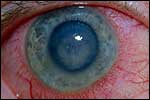
Neovascularization following stromal keratitis is often a sign of chronic, recurrent disease. Less commonly, neovascularization can progress more rapidly during an episode of severe inflammation. Vessels in herpes simplex stromal keratitis can be superficial, mid-stromal, deep, or at multiple layers. Inflammation in the presence of neovascularization can lead to lipid deposition, which can be visually debilitating. Active vessels will also decrease the success of corneal transplantation. Aggressively treated inflammation can limit the growth of new vessels and is essential in preventing severe visual loss. Longstanding quiescent disease can lead to ghost vessel formation.
Endotheliitis, commonly called keratouveitis, may occur with or without overlying stromal edema. The endothelium is typically affected in a disciform pattern, but the presentation may be diffuse or linear. Keratic precipitates, which may be granulomatous in appearance, are commonly present, but may be absent. The granulomatous nature of these precipitates, as well as the presence of viral antigens, has been confirmed histopathologically. When arranged in a linear manner along the endothelium in a corneal transplant, keratic precipitates can mimic a Khodadoust line.6 In some cases, the difference can be detected by observing that the HSV-induced reaction extends onto the host bed.
Iridocyclitis may accompany any of the above forms of keratitis or may occur independently without any previous history of ocular HSV. An anterior chamber reaction, keratic precipitates, and an increased intraocular pressure due to a concomitant trabeculitis are common features. Increased eye pressure in the presence of idiopathic iridocyclitis should raise the suspicion of herpetic disease. The precipitates may be granulomatous and may extend above the midline (outside Arlt’s triangle). Posterior synechiae may form in herpetic iridocyclitis, and hyphema is a rare but reported finding. Presence of a hypopyon is almost always limited to necrotizing stromal keratitis and is usually rare. Usually, hypopyon signals secondary infection with another organism.3 Segmental iris atrophy and transillumination defects may indicate previous episodes of herpetic iridocyclitis.
Acute retinal necrosis and progressive outer retinal necrosis are potentially devastating disorders caused by herpes family viruses and characterized by acute confluent peripheral retinitis, papillitis, and anterior chamber reaction. It is rare for herpes keratitis to be associated with either acute retinal necrosis or progressive outer retinal necrosis. Although dilated fundoscopy is a part of any complete examination, screening for these conditions in the setting of herpes keratitis is unlikely to be rewarding.7
Laboratory Diagnosis
Diagnostic scraping for microbiologic confirmation in suspected herpes simplex keratitis is typically reserved for problematic cases, such as necrotizing stromal keratitis. Most cases of herpetic keratitis can be diagnosed on clinical grounds. If viral cultures are taken, results may be delayed for several weeks. Immunologically based tests such as the Herpcheck or ELISA testing may produce results within hours rather than days or weeks.
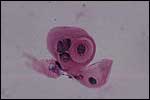
Cytologic examination of Giemsa- or Wright-stained specimens may reveal multinucleated giant cells and the more pathognomonic Cowdry type A inclusions that are found with HSV infection (Slide 4). Immunohistochemical staining specific for HSV-1 or HSV-2 may also be useful. Such investigations are usually reserved for corneal biopsies or for examination following lamellar or penetrating keratoplasty in eyes with recurrent or persistent scarring of unknown cause.
The use of serologic antibody tests (IgM + IgG for HSV) is controversial. Antibody titers can fluctuate independently of the presence of disease, and low positive titers are nearly ubiquitous in the adult population due to the widespread nature of herpetic disease. Both false-positive and false-negative test results are common. Studies examining recrudescence of HSV-1 have found no clear association with IgG titers.8 However, in clinical practice, a suspicious flareup of keratitis associated with high HSV titers, along with a lower titer obtained during a period of inactive disease, can be considered strongly suggestive of HSV as an etiologic agent.
Treatment
Medical treatment for herpes simplex keratitis consists of antiviral and corticosteroid therapy, both of which can be delivered topically or orally.
Antivirals The antivirals most commonly used in the United States include topical idoxuridine (IDU), vidarabine and trifluridine, and oral acyclovir, valacylcovir, and famcyclovir. All of these antivirals limit viral replication by interfering with protein synthesis, and are thus virostatic. Because they are not virocidal, they cannot eliminate latent infections.
IDU, which was originally used as an anticancer drug, is a thymidine analog that can be incorporated into viral RNA, leading to the production of nonsense proteins. IDU can also bind to and inhibit viral DNA polymerase. Because IDU can be incorporated into host DNA, it has a relatively high toxicity in topical form. In the treatment of HSV epithelial keratitis, IDU has been found to be less effective than the other antivirals discussed below.9 It is available as a 0.1% topical solution.10
Trifluridine, or trifluorothymidine, also originally developed as an anticancer drug, has a similar method of action and a similar side effect profile to that of IDU. Like IDU, trifluridine results in considerable toxicity in topical form, producing a superficial punctate keratitis and other signs. Its topical form is available as a 1% solution.
Vidarabine (Ara-A) is converted into an active form by host cellular enzymes. It works by inhibiting viral DNA polymerase, acts as a DNA chain terminator, and can also be incorporated into viral (and host) DNA. Its incorporation into host DNA is limited, so it has a much lower side effect profile than IDU. Vidarabine has a low solubility and the topical medication is only available as a 3% ointment, which may be preferable to drops in some patients (e.g., children). Use of oral vidarabine is primarily limited to cases in which viral resistance to more potent antivirals (acyclovir, famcyclovir, and valacyclovir) is suspected.
Acyclovir, as well as valacylcovir and famcyclovir, has specific activity against HSV. Thus, these agents can spare host tissues and exhibit considerably lower toxicity than IDU or vidarabine. These drugs achieve their specific activity against HSV by requiring initial activation (phosphorylation) by a viral-specific thymidine kinase. Once converted to the monophosphate form by viral thymidine kinase, the drugs are then converted to a triphosphate form by the cell and undergo additional modifications.
Acyclovir is a purine nucleoside. The active drug becomes incorporated into viral DNA and forms an inactive and irreversible complex between DNA and DNA polymerase, thus terminating synthesis in a process known as obligate chain termination.11 Immunocompromised patients under long-term treatment may develop resistance to acyclovir due to a mutation in viral thymidine kinase.10 Such HSV strains should be considered to be resistant to valacyclovir and famcyclovir and may also be resistant to trifluridine, which requires viral thymidine kinase for activation. Acyclovir is available only as an oral agent in the United States, but a topical form is available in Europe and elsewhere.
Valacyclovir is the L-valine ester of acyclovir. The addition of the salt moiety greatly increases bioavailability, allowing lower dosing at less frequent intervals. Valacyclovir administration has been found to result in plasma acyclovir levels that are three to five times higher than that of high-dose acyclovir.12 In our practice, valacyclovir is the oral antiviral of choice. Although more expensive than acyclovir, its higher bioavailability and longer dose intervals may result in better compliance and improved outcomes.
Famcyclovir is a prodrug of penciclovir, which is a guanosine analog. Once activated by viral thymidine kinase and processed by the host cell, it acts as a competitive inhibitor of guanosine during viral DNA polymerization, thus leading to unusable proteins.
Management
Treatment of HSV keratitis is divided into different modalities, similar to the approach to diagnosis as described above.
Epithelial dendritic keratitis can be treated with debridement, topical antiviral therapy, oral antiviral therapy, or a combination of the above. Debridement is often best performed gently either by rolling (rather than rubbing) a cotton swab over the affected area or by placing and then removing a strip of filter paper placed over the lesion. By minimizing trauma during debridement, one can limit trauma to surrounding cells, decrease the risk of releasing viral particles from infected cells, and decrease the risk of mechanically pushing viral particles deeper into the cornea. Topical antiviral therapy is typically used in much the same way as topical antibiotic therapy: intensive topical therapy (either trifluridine 1% solution eight times a day, or vidarabine 3% ointment five times a day), tapering to lower doses after 5 to 7 days (trifluridine four times a day, vidarabine three times a day). Therapy should be stopped in 10 to 14 days unless there is evidence of persistent dendritic epithelial keratitis. It should be noted that such persistence is extremely rare. It is more common to see persistent epithelial defects due to drug toxicity, a neurotrophic condition, superinfection with another agent, or misdiagnosis. Particularly, persistent dendriform defects in a contact lens wearer, especially if the cornea is neurotrophic, should raise the suspicion of Acanthamoeba keratitis.
Oral antiviral therapy should always be considered in the treatment of epithelial herpes simplex keratitis. Oral acyclovir has been shown to be as effective as topical agents against epithelial herpes simplex keratitis, and the lower topical side effect profile of these agents makes them useful in selected patients, including children.13,14
Epithelial geographic keratitis can be treated with oral and topical antiviral therapy to speed the reduction of topical therapy and promote healing. Such lesions can also be treated with a topical antibiotic drop as prophylaxis against bacterial superinfections.
Neurotrophic keratitis is best treated with modalities that are aimed at keeping the corneal surface moist and well covered. Artificial tears, punctal plugs, and topical emollients can be considered first-line therapy in this disorder, although their success is likely to be limited. Temporary contact lenses, used with prophylactic topical antibiotic drops (one to four times a day), can result in improvement of small lesions. For large lesions or for patients who are not considered good contact lens candidates, tarsorrhaphy with tape, glue, or surgery provides definitive management. A conjunctival flap may be necessary when these measures are inadequate.
Because necrotizing stromal keratitis is marked by the presence of active viral particles, treatment with oral antiviral therapy is essential. My colleagues and I have found that high doses of antivirals (e.g., valacyclovir 1 g three times a day) are well-tolerated and effective in reducing the intensity of inflammation in these cases and allowing healing of any overlying epithelial defects. The difficulty in managing necrotizing disease is in balancing antiviral and anti-inflammatory therapy. We recommend judicious use of topical steroids following high-dose administration of antivirals. One study has found topical cyclosporine A to be of benefit in these cases instead of topical steroids.15
Disciform stromal keratitis has been found to respond positively to the application of topical corticosteroids (prednisolone phosphate) in the presence of concomitant topical antiviral therapy. The Herpetic Eye Disease Study (HEDS) established that this regimen resulted in a reduction of both the intensity and duration of stromal inflammation.16 We prefer to use more potent topical corticosteroids, along with oral (rather than topical) antiviral therapy. The literature supports this approach; one HEDS paper looking at stromal keratitis treated with mild steroids and topical antivirals found a benefit in 6-month visual acuity in patients who also received oral antivirals (acyclovir 2 g per day). Oral antivirals have also been shown to effectively treat and prevent epithelial keratitis and to decrease the risk of recurrent stromal keratitis.1,13
The HEDS study found a trend toward improved response of herpetic iridocyclitis and trabeculitis when oral acyclovir was added to topical trifluridine and corticosteroids. A combination of oral antivirals and topical corticosteroids may provide sufficient antiviral coverage to allow for the desired suppression of immune response.
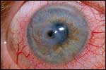
Penetrating keratoplasty (PK) is frequently performed after visually significant herpetic corneal scarring and/or descemetocoele formation (Slide 5). Relative to PK for keratoconus or mild bullous keratopathy, PK following herpetic disease is accompanied by an increased risk of graft failure and recurrence of herpetic disease. Prophylaxis, as described below, should be routinely used.
Prevention
The HEDS study has established that long-term (12-month) oral antiviral therapy, with a dose of acyclovir 400 mg twice a day, can reduce the recurrence of epithelial keratitis and stromal keratitis.1 The benefit of such therapy was found to be greatest among patients with previous stromal keratitis, but in all groups benefits were found even if the previous episode of eye disease had occurred up to a year before therapy. For patients with epithelial keratitis only, the decision to begin prophylactic therapy must be made on an individual basis, but typically such prophylaxis is reserved for patients with two or more episodes in a year. In the treatment of epithelial keratitis, the HEDS study group did not find a benefit to adding 3 weeks of intensive oral acyclovir therapy to topical trifluridine.17
Patients with any previous herpes simplex keratopathy who are undergoing PK may benefit from both preoperative and postoperative prophylaxis, and we do this routinely.18 Similar prophylaxis is much more important for patients with suspected herpetic eye disease undergoing PRK or LASIK, since the short-wavelength laser can reactivate herpetic keratitis and lead to significant morbidity.19-21
References
-
The Herpetic Eye Disease Study Group. Oral acyclovir for herpes simplex virus eye disease: Effect on prevention of epithelial keratitis and stromal keratitis. Arch Ophthalmol. 2000;188(8):1030-1036.
-
Liesegang TJ, Melton J, Daly PJ, et al. Epidemiology of ocular herpes simplex. Arch Ophthalmol. 1989;107:1155-1159.
-
Boisjoly HM, Pavan-Langston D, Kenyon KR, et al. Superinfections in herpes simplex keratitis. Am J Ophthalmol. 1983;96(3):354-61.
-
Pepose JS. Herpes simplex keratitis: Role of viral infection versus immune response. Surv Ophthalmol. 1991;35:345-352.
-
Schwartz GS, Harrison AR, Holland EJ. Etiology of immune stromal (interstitial) keratitis. Cornea. 1998;17(3):278-281.
-
Holbach LM, Font RL, Naumann GO. Herpes simplex stromal and endothelial keratitis. Granulomatous cell reactions at the level of Descemet’s membrane, the stroma, and Bowman’s layer. Ophthalmology 1990;97(6):722-728.
-
Sado K, Kimura T, Hotta Y, et al. Acute retinal necrosis syndrome associated with herpes simplex keratitis. Retina. 1994;14(3):260-263.
-
Doerr HW, Rentschler M, Scheifler G. Serologic detection of active infections with human herpes viruses (CMV, EBV, HSV, VZV): Diagnostic potential of IgA class and IgG subclass-specific antibodies. Infection. 1987;15(2):93-98.
-
Wilhelmus KR. The treatment of herpes simplex virus epithelial keratitis. Trans Am Ophthalmol Soc. 2000;98:505-532
-
Merck Manual Online (www.merck.com/pubs/mmanual/section13/chapter154/154a.htm, 5/20/2002).
-
Furman PA, St Clair MH, Spector T. Acyclovir triphosphate is a suicide inactivator of the herpes simplex virus DNA polymerase. J Biol Chem. 1984;259(15):9575-9579.
-
Weller S, Blum MR, Doucette M, et al. Pharmacokinetics of the acyclovir pro-drug valacyclovir after escalating single- and multiple-dose administration to normal volunteers. Clin Pharmacol Ther. 1993;54(6):595-605.
-
Collum LMT, Akhtar J, McGettrick P. Oral acyclovir in herpetic keratitis. Trans Ophthalmol Soc UK. 1985;104:629-632.
-
Schwartz GS, Holland EJ. Oral acyclovir for the management of herpes simplex virus keratitis in children. Ophthalmology. 2000;107(2):278-282.
-
Heiligenhaus A, Steuhl KP. Treatment of HSV-1 stromal keratitis with topical cyclosporin A: A pilot study. Graefe’s Arch Clin Exp Ophthalmol. 1999;237(5):435-438.
-
Wilhelmus KR, et al. Herpetic eye disease study, a controlled trial of topical corticosteroids for herpes simplex stromal keratitis. Ophthalmology. 1994;101:1883-1896.
-
The Herpetic Eye Disease Study Group. A controlled trial of oral acyclovir for the prevention of stromal keratitis or iritis in patients with herpes simplex virus epithelial keratitis. Arch Ophthalmol. 1997;115(6):703-712.
-
Barney NP, Foster CS. A prospective randomized trial of oral acyclovir after penetrating keratoplasty for herpes simplex keratitis. Cornea. 1994;13:232-236.
-
Perry HD, Donnenfeld ED, Doshi SD, et al. Perforated corneal ulcer following LASIK. CLAO. 2002;28:4:69-71.
Bibliography
Barron BA, Gee L, Hauck WW, et al. Herpetic Eye Disease Study. A controlled trial of oral acyclovir for herpes simplex stromal keratitis. Ophthalmology. 1994;101(12):1871-82.
Holland EJ, Mozayeni RM, Schwartz GS. Herpes Simplex Keratitis. In: Cornea; Krachmer JH, Mannis MJ, Holland EJ (eds.) Mosby 1997.
Holland EJ; Schwartz GS. Classification of herpes simplex virus keratitis. Cornea 1999 Mar; 18(2):144-54.