Introduction
Complex clinical problems in corneal and external disease often leave both the patient and the physician perplexed. I have found that cytology may be of benefit in helping to narrow the differential diagnosis.1 In some cases, it will even point to a specific etiology, resulting in a new treatment plan to effectively cure the patient. Cytology also may allow for a more rapid diagnosis than culture, as culture may be delayed up to several weeks in cases of opportunistic organisms and fungi.
In order to make cytology work for you, there is some required equipment needed. The first item is a Kimura spatula with a pointed edge, a Storz E-1090. Second, a Coplin jar is needed. A Coplin jar is scored on the inside and allows up to five separate glass slides to be stored. Thirdly, methyl alcohol is required. And last, cytology slides will be needed. These slides have a small white circle in the center for placement of the scrapings. Once the scraping has been placed in the white circle on the slide, it is immediately placed into the Coplin jar with methyl alcohol. The smears are fixed instantaneously, and staining may take place immediately or even up to several weeks later. The jar may be sent to a local hospital to be processed and evaluated. An expert cytologist will charge as little as $50 to evaluate and process a patient's specimen.
For physicians who want immediate answers, Gram and Giemsa stain kits are available from Harco. Gram stains are good for bacteria, fungi, and Acanthamoeba but are only fair for cytology. Giemsa is good for viral inclusions and general cytology. It may also be modified for eosinophyls and basophiles. Commonly, both Gram and Giemsa smears are needed, although Papinicolau may be substituted for Giemsa. Gomori Methanamine Silver or Periodic Acid Schiff are best for fungi. Calcofluor white is the best stain for Acanthamoeba.
Indications
Conjunctivitis
In differentiating among bacterial,
viral, and allergic disease, note that polymorphonuclear leucocytes are
characteristically seen in bacterial infections while mononuclear cells are
seen in viral disease. Eosinophils and basophils are characteristic of allergic
disease.
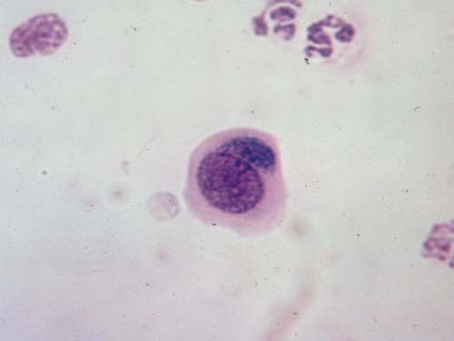
Evaluating chronic cases for cytology and inclusions also can be helpful. An example is MK, a 56-year-old black woman who was treated for 2 years for superior limbic keratoconjunctivitis. A conjunctival scraping showed inclusions (Halberstaedter-von Prowazek) characteristic of inclusion conjunctivitis of adults (Slide 1). The patient was treated with systemic tetracycline and within 1 week was totally asymptomatic and showed resolution of her chronic keratoconjunctivitis.2
Keratitis
Fluoroquinolones have virtually
eliminated the need for cultures and corneal scrapings. However, in cases with
central corneal ulcers and hypopyon, cultures and scrapings are in a patient's
best interests. Corneal scrapings are very important in making the diagnosis of
fungal and Acanthamoeba infections. Smears alone will make the diagnosis in 85%
of fungal infections and 92% of Acanthamoeba cases.3 Investigation
of cases with chronic superficial keratitis may also prove useful to the
clinician.
A patient presented to me with bilateral superficial keratitis, decreasing her vision to 20/200 in both eyes. Numerous topical agents were used, including several antibiotics and corticosteroids. A superficial scraping showed epithelial cells with eosinophilic intranuclear inclusions (Cowdri type A) and giant cells (Slide 2). Treatment with topical Viroptic (trifluridine, Glaxo) quickly resulted in a cure and a return of normal vision.
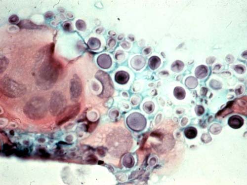
In corneal transplants, scrapings of any infiltrates are often helpful. One patient presented at 8 months posoperatively with a small superficial epithelial and subepithelial infiltrate (Slide 3). This lesion was smeared and cultured. The smear showed yeast of varying diameters with thick walls characteristic of Cryptococcus (Slide 4). This diagnosis was confirmed by cultures, and the patient quickly responded to topical miconazole (10 mg/mL).4
Lids
Smears of chronic blepharitis will show
bacteria or occasionally parasites and, rarely, malignant cells. Confirmation
of a clinical diagnosis such as Molluscum contagiosum may be easily made by
cytology.5
Summary
The performance of smears may be of help to the busy clinician in cases in which usual therapeutic approaches have failed or when clinical suspicion suggests performance of this test will benefit the patient.
References
-
Perry HD, Nauheim JS, Donnenfeld ED. Nocardia asteroids keratitis presenting as a persistent epithelial defect. Cornea. 1989;8(1):41-44.
-
Seedor JA, Droste P, Perry HD. Chlamydial infection producing keratoconjunctivitis of the corneoscleral limbus. Am J Ophthalmol. 1986;102:6:798-800.
-
Perry HD, Donnenfeld ED. Cryptococcal keratitis post-keratoplasty. Am J Ophthalmol. 1990;110(3):320-321.
-
Wilhelmus KR, Osato MS, Font RL et al. Rapid diagnosis of Acanthamoeba keratitis using Calcofluor white. Arch Ophthalmol. 1986;104:1309.