Introduction
Meyer-Schwickerath has been credited with early attempts at retinal photocoagulation; first with sunlight in 1946, then with his development of the xenon-arc lamp in 1956.1 The first ophthalmic laser was developed by Maiman in 1960, utilizing the ruby crystal. 2 The LASER (light amplification by the stimulated emission of radiation) has the advantage of monochromatic light that is spatially and temporally coherent. Since their inception, lasers have been valuable in the treatment of a wide variety of medical conditions. Previously untreatable disease entities have subsequently done well with the new laser techniques. Ophthalmic laser investigation has coincided with the development of new laser technology. Since the introduction of the ruby red laser, other lasers have come to play a more important role in the treatment of retinal disorders.3
Laser Types and Wavelengths (Slide 1)
The argon blue-green laser (488 nanometers [nm] and 514 nm) was introduced by L’Esperance in 1968.4 This was the first laser that was well absorbed by hemoglobin, a quality of great importance when treating vascular lesions directly. Unfortunately, the blue component is also highly absorbed by xanthophyll, causing undesired inner retinal tissue damage during macular laser photocoagulation. This absorption quality has led to the argon blue-green laser being contraindicated for the use of macular laser photocoagulation. It has also been shown that long-term use of the blue wavelength may decrease color discrimination in ophthalmologists who treat these patients.5
Argon green laser (514 nm) is minimally absorbed by xanthophyll, with higher absorption by both hemoglobin and melanin. These absorption qualities make argon green a much safer alternative for macular photocoagulation and the most popular wavelength for retinal photocoagulation in general.
Krypton red laser (647 nm), developed by Laser Tek (Finland) in 1978,6 is absorbed essentially by melanin only. The red wavelength is an excellent choice for macular laser photocoagulation, with no absorption by xanthophyll. It is also effective for treating through media opacities such as cataracts and vitreous blood. Treatment of choroidal neovascularization with overlying blood is possible with this wavelength with less chance of potential damage to the inner retinal layers. Eyes with less pigment may require higher energy levels, or may not be able to produce the desired effect. Other disadvantages include the inability to treat vascular lesions, the inability to directly coagulate vessels in the event of an inadvertent hemorrhage, and the greater likelihood of discomfort due to the deeper choroidal penetration.
In 1984, L’Esperance offered another option for laser photocoagulation with the development of the tunable dye lasers (570 nm to 630 nm). 7 The dye lasers use multiple dyes to produce emissions over a wide range of wavelengths. Theoretically, this allows the operator to select the desired wavelength to fit the specific needs of the procedure being performed. Dye yellow at 577 nm is not well absorbed by macular xanthophyll and is extremely well absorbed by hemoglobin. These qualities make this wavelength ideal for the treatment of vascular anomalies, as well as choroidal neovascular membranes. Dye yellow, however, is not as effective as the other wavelengths when treating through vitreous or preretinal blood. Dye red laser (630 nm) is a useful alternative to krypton red. Other dye wavelengths may have theoretical advantages, yet little clinical utility. The major disadvantage of dye lasers is the cumbersome and complex quality of these laser systems, with the dyes prone to degradation, requiring frequent maintenance.
The diode laser (810 nm), developed in 1987, emits radiation with excellent electrical-to-optical efficiency. These lasers have become popular due to their low cost, portability, standard electrical requirements, high efficiency, and lack of water-cooling system requirements. A disadvantage of this wavelength is the deeper penetration of the near-infrared light with a greater likelihood of pain, and less visible burns. The diode wavelength lies at the peak of the absorption spectrum of indocyanine green dye, and may play a role in the selective ablation of choroidal neovascularization. A more recent procedure is photodynamic therapy (PDT), which involves the use of a photosensitizer and the diode laser. The photosensitive dye is injected systemically and allowed to accumulate in the more metabolically active proliferating tissues. The diode laser, at the correct wavelength, activates these molecules to generate singlet oxygen and free radicals, which result in damage to the cells that contain the dye.
The Nd:YAG laser (1,064 nm) penetrates deeper than the other lasers due to the lower absorption by melanin. A potassium-titanium-phosphate (KTP) crystal has been used to double the frequency and halve the wavelength of the Nd:YAG laser. The resultant laser is a yellow-green laser at 532-nm wavelength. This double-frequency Nd:YAG laser is ideal for macular laser, due to the lower xanthophyll and higher hemoglobin and RPE absorption. It also offers the dependability and low maintenance of a solid-state, air-cooled laser.
Retinal Laser Use
Informed consent is necessary for any procedure that is to be performed on a patient. The discussion should include the choice of anesthesia, ranging from topical, to retrobulbar or peribulbar infiltration, to general anesthesia. If akinesia is required for the procedure, then one of the infiltrative alternatives should be chosen. The procedure is usually office-based, with the occasional necessity for the operating room if the patient requires a general anesthetic. Slit lamp and mounted indirect ophthalmoscope lasers may be used in the office on an outpatient basis. There are a variety of contact lenses available for the slit lamp laser (Slide 2 and Slide 3). These range from the universal three-mirror Goldmann contact lens, to the Peyman fundus contact lens, to the inverted image system lenses by Rodenstock, Mainster, and more recently, Volk’s QuadrAsperic fundus lens and the Super Quad 160 fundus laser lens.
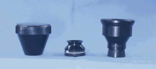
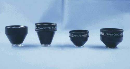
The choice of laser apparatus, wavelength, and laser settings is dependent on the type of laser procedure to be performed.
Basic Complications
Laser photocoagulation is generally thought to be a less invasive, more conservative modality in the field of vitreoretinal surgery. Yet there are definite risks and potential complications that the surgeon should consider prior to treatment.8
Pain is not a common complication of macular laser. Pain can occur with longer duration laser settings for subretinal neovascularization, and the longer wavelength lasers (red or diode). More typical is the discomfort experienced during peripheral panretinal photocoagulation, which often necessitates an infiltrative anesthetic. Post laser pain can be the residual discomfort from the laser itself, or the occasional postoperative corneal abrasion secondary to the contact lens. Breaks in Bruch’s membrane are more likely to occur with intense laser burns produced with a smaller spot size at a shorter duration.9 This can become manifest by hemorrhage, with the subsequent development of choroidal neovascularization. The neovascularization can bleed subretinally, or extend into the subhyaloid space with vitreous hemorrhage, or traction retinal detachment.
Choroidal hemorrhages can also occur with laser settings similar to those that produce breaks in Bruch membrane. This can have serious consequences with poorer visual prognosis if a hemorrhage occurs while treating a choroidal neovascular membrane. The hemorrhage tends to arise in a nonphotocoagulated area of the net, thus explaining the logic behind the strategy of lasering the foveal edge of the membrane first, to minimize the risk of subfoveal hemorrhage.
More intense laser and higher total shots during panretinal photocoagulation increase the risk of exudative choroidal detachments. With significant choroidal detachments, the iris-lens diaphragm may shift forward with the development of a secondary angle closure glaucoma. Panretinal photocoagulation should thus be divided into multiple sessions, with moderate laser intensity.
Iatrogenic retinal pigment epithelial tears can occur during or after the treatment of choroidal neovascularization, more commonly with an associated pigment epithelial detachment. The thermal effect of the laser on the choroidal vasculature can secondarily result in vascular contraction and traction on the overlying RPE. This can have a variable result on the vision, ranging from no change in vision, to severe visual loss.
Macular edema can occur in diabetic eyes after intense panretinal photocoagulation. This is thought to be caused by an alteration in the hemodynamic equilibrium within an already compromised retinal vasculature. This can possibly be avoided by treating any pre-existing diabetic macular edema first, with subsequent PRP to follow. Often, the macular edema is temporary, with resolution over the following few weeks.
Epiretinal membrane proliferation or macular pucker with resultant metamorphopsia and visual loss can occur after laser photocoagulation. Again, this is more common with intense laser burns. The dissipated heat from the inner retina can cause damage to the internal limiting membrane (ILM). Small holes in the ILM can lead to macrophage, fibroblast, and RPE migration with subsequent membrane formation.
Visual field loss and reduction in night vision are common occurrences after extensive panretinal photocoagulation. These were more significant in the early days with the use of the xenon-arc photocoagulator.
Accidental foveal burn, though infrequent, can be a disastrous complication with marked visual loss. This can occur during macular laser with poor patient cooperation, or inadequate akinesia and anesthesia. This can also occur during PRP secondary to the surgeon not being aware of the location being treated; either through a peripheral mirror, accidentally slipping to the central view, or inexperience with the inverted lens system.
Laser Tissue Interactions
The indications of laser photocoagulation to the retina have expanded dramatically since the early days of Meyer-Schwickerath. A complete all-encompassing description of the indications is beyond the scope of this discussion. Only the more common entities will be included, with mention of other entities, which have similar endpoint pathologic components.
Diabetic retinopathy was the first indication for laser photocoagulation, and remains the most commonly treated entity. There have been several randomized, prospective clinical trials documenting the efficacy of laser photocoagulation in the treatment of diabetic retinopathy. The first study, the Diabetic Retinopathy Study (DRS), began in 1971 under the sponsorship of the National Eye Institute.10,11 The DRS established the efficacy of panretinal laser photocoagulation for the treatment of proliferative diabetic retinopathy. The investigators identified certain findings as risk factors for severe visual loss.12 These consisted of:
- New vessels
- New vessels on or within 1 disc diameter (DD) of the optic disc (NVD)
- Severity of the new vessels
- Vitreous or preretinal hemorrhage
Three or more of these risk factors carried a significantly higher chance of severe visual loss, defining the high-risk characteristics necessitating treatment.
The treatment protocol consists of scatter laser, originally performed with xenon arc or argon blue-green, now most commonly with argon green laser. The laser spots are spaced one half to one spot size apart, with a 500-µm spot size, a few thousand spots per eye. This is placed outside the arcades, extending to the peripheral retina, divided into two to three separate laser sessions.
Diabetic macular edema is the most common manifestation of diabetic retinopathy, and the most common cause of visual loss in the diabetic population. The Early Treatment Diabetic Retinopathy Study (ETDRS), a National Eye Institute-supported randomized multicenter clinical study, definitively established, among other things, the effectiveness of focal or grid laser photocoagulation for clinically significant diabetic macular edema (CSME).13 The study defined CSME as one of the following:
- Retinal thickening at or within 500 µm of the center of the macula
- Hard exudates at or within 500 µm of the center of the macula associated with adjacent retinal thickening
- Retinal thickening of 1 DD or greater within 1 DD of the center of the macula
Microaneurysms ranging from 500 µm to 3,000 µm from the center of the macula should be treated with focal laser. The ETDRS recommended 50-µm to 100-µm spot sizes, exposure time 0.1 second or less with moderate intensity (Slide 4). Retreatment was considered if CSME persisted at the 3 to 4 month follow-up, with microaneurysms up to 300 µm from the center of the macula considered to be treatable.14
Branch retinal vein occlusion (BRVO) is a very common retinal vascular disorder, second only to the more commonly occurring diabetic retinopathy.15 In the natural history of BRVOs, reduction of visual acuity can occur for a variety of reasons. Acutely, the presence of dense intraretinal hemorrhages with occasional preretinal blood can lead to a reduction of vision. Chronically, macular pathology from macular edema or macular ischemia can both result in visual loss. Retinal ischemia can also cause secondary retinal neovascularization with the development of vitreous hemorrhage and traction retinal detachment.
Initial studies on laser photocoagulation of BRVOs often vary in their recommendations.16-18 A prospective, randomized, controlled clinical trial was required to establish the natural course of the disease, and the efficacy of treatment.15,19 The Branch Vein Occlusion Study Group was established to answer the following questions:
- Could peripheral scatter argon-laser photocoagulation prevent the development of neovascularization?
- Could peripheral scatter argon-laser photocoagulation prevent vitreous hemorrhage?
- Could macular argon-laser photocoagulation improve visual acuity in eyes with macular edema reducing vision to 20/40 or worse?
The conclusions of the study led to the following clinical recommendations:
For vision reduced to 20/40 or worse:
- Wait for sufficient clearing of retinal hemorrhage to obtain high-quality fluorescein angiography.
- Evaluate fluorescein angiography for macular edema versus macular nonperfusion.
- If macular edema explains visual loss, and vision continues to be 20/40 or worse without spontaneous improvement after 3 months of observation, grid-pattern macular photocoagulation is recommended.
- If macular nonperfusion explains visual loss, no treatment is available to improve vision.
For retinal involvement area greater than 5 DD in size:
- Wait for sufficient clearing of retinal hemorrhage to obtain high-quality fluorescein angiography.
- Evaluate fluorescein angiography for nonperfusion. If there are more than 5 DD of nonperfusion, follow-up at 4-month intervals for development of neovascularization.
- If neovascularization develops and is confirmed, perform laser photocoagulation in the involved quadrant according to protocol described.
Grid laser photocoagulation of macular edema consists of numerous medium white laser burns over the area of capillary leakage as identified on the fluorescein angiogram. A 100-µm laser spot size is utilized, extending no closer than the edge of the foveal avascular zone and no more peripheral than the respective vascular arcade. The treated eye is reevaluated at 4 months with repeat fluorescein angiography. The clinical exam, along with the angiogram results, determines the need for further laser.
Laser photocoagulation for retinal neovascularization complicating a BRVO consists of peripheral scatter laser involving the entire area of involved retina (Slide 5). A 200-µm to 500-µm laser spot size is utilized, extending no closer than 2 DD to the center of the fovea. Repeat laser photocoagulation should be considered for persistent neovascularization, or the development of a vitreous hemorrhage.
Another vaso-occlusive disorder, central retinal vein occlusion (CRVO), can cause significant visual loss. CRVOs can be divided into nonischemic or perfused, and ischemic or nonperfused. Both types can cause diffuse capillary leakage with marked macular edema. The ischemic type can also lead to neovascularization with possible anterior segment involvement and neovascular glaucoma, or posterior segment involvement and vitreous hemorrhage. The treatment of CRVOs and their complications has been somewhat controversial in the past. For this reason, the Central Vein Occlusion Study (CVOS) was designed to answer the question of when to intervene in CRVO patients with complications.20,21 Nine participating clinical centers enrolled 728 eyes of 725 patients into the CVOS. The following treatment recommendations were made from analysis of the study data:
- Macular grid-pattern laser photocoagulation for eyes with perfused macular edema is currently not recommended.
- Prophylactic panretinal photocoagulation, before the development of iris or angle neovascularization (INV/ANV), does not prevent the development of INV/ANV in all eyes and is not recommended. Prompt regression of neovascularization, in response to panretinal photocoagulation, is more likely to occur in those eyes that have not previously received panretinal photocoagulation.
- Although the visual prognosis is guarded and depends upon the initial status, careful monitoring for INV/ANV and prompt panretinal photocoagulation treatment when early INV/ANV appears should diminish the development of neovascular glaucoma.
Choroidal neovascularization (CNV) is a common pathologic condition caused by a wide variety of clinical entities, the most common being age-related macular degeneration (AMD). Other causes of CNV include presumed ocular histoplasmosis syndrome (POHS), myopic degeneration, angioid streaks, choroidal ruptures, tumors, radiation retinopathy, idiopathic, and others. A detailed discussion of the indications for laser photocoagulation of all these conditions with CNV is beyond the scope of this tutorial. The more common entities will be described briefly.
AMD is the most common cause of blindness in the United States and other developed countries. The exudative or neovascular form of AMD accounts for the majority of cases of severe visual loss, yet is less common then the nonexudative or dry form of AMD. The Macular Photocoagulation Study (MPS) Group was originally constructed to answer the question of whether laser photocoagulation was effective for the treatment of CNV complicating AMD.22 They subsequently looked at patients with POHS and idiopathic CNV as well.23,24 The MPS classified CNV as extrafoveal, juxtafoveal, and subfoveal according to the location of the CNV in relation to the fovea. (Slide 6) For AMD, POHS, and idiopathic groups with extrafoveal or juxtafoveal lesions, laser-treated eyes lost less vision than untreated eyes, with high persistence and recurrence rates. For subfoveal lesions, only AMD patients were considered, with an initial decrease in visual acuity noted in treated eyes, with some benefit demonstrated at the 2-year follow-up.25
Extrafoveal CNV, defined as a well-demarcated CNV 200 µm to 2,500 µm from the center of the foveal avascular zone (FAZ), may be treated with or without retrobulbar anesthesia. The neovascular complex is treated in a confluent fashion to produce uniform retinal whitening (Slide 7). Treatment of the lesion extends 100 µm beyond the borders, using argon green laser 200-µm to 500-µm spot size at 0.1 seconds to 0.5 seconds duration. Peripapillary CNV may be treated if the laser photocoagulation will spare at least one and one-half hours of the peripapillary nerve fiber layer temporally.
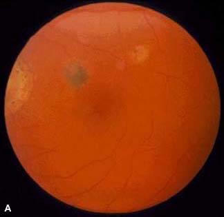
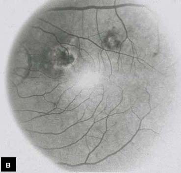
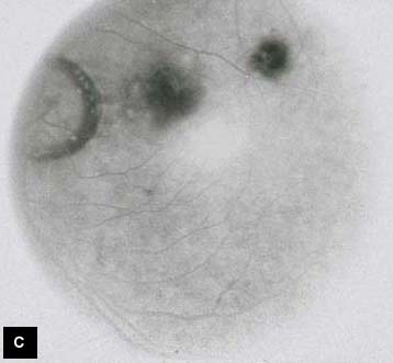
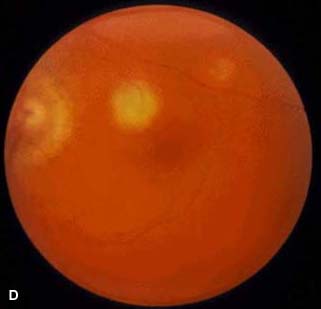
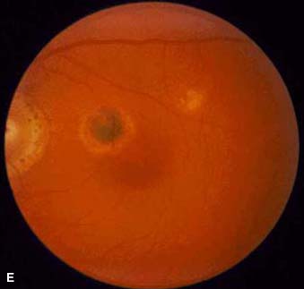
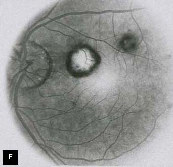
Juxtafoveal CNV is defined as a well-demarcated CNV 1 µm to 199 µm from the center of the FAZ. The CNV may also be more than 200 µm from the center of the FAZ with adjacent blood or pigment extending within 200 µm. The MPS investigators recommended retrobulbar anesthesia with krypton red laser. Again, the neovascular complex is treated in a confluent fashion to produce uniform retinal whitening. A 100-µm margin is recommended, except on the foveal side, where treatment beyond the borders of the lesion is dependent on the distance the hyperfluorescence is from the center of the FAZ.
Subfoveal CNV is defined as a well-demarcated CNV that extends under the center of the FAZ. With the advent of photodynamic therapy (PDT), it is difficult to justify conventional thermal laser photocoagulation of subfoveal lesions. There now is very good data supporting the efficacy of PDT with Visudyne (Novartis Ophthalmics) for the treatment of subfoveal lesions secondary to AMD and myopic degeneration. PDT requires the infusion of a photosensitizer dye that accumulates in rapidly proliferating tissue such as choroidal neovascularization. The neovascular tissue is then irradiated with light of a wavelength that coincides with the absorption peak of the dye. For Visudyne dye, this would consist of the diode laser with a wavelength of 690 nm. This initiates a photochemical reaction with free radical and singlet oxygen production, which promotes vascular occlusion of the neovascular tissue. Revascularization occurs over the ensuing three months with repeat treatment performed every three months until no further leakage remains on fluorescein angiography.26
Prophylactic treatment of retinal breaks is somewhat more controversial. The indications for such treatment vary depending on the reference source. Most specialists would agree with the treatment of symptomatic horseshoe (flap) tears. From 25% to 90% of these tears have been found to lead to retinal detachments. Prophylactic treatment can reduce this incidence down to 0% to 19%.27 Treatment consists of a triple row of nonconfluent laser burns of moderate to heavy intensity, 200 µm, and 0.1 second duration.
Other treatable entities, though not as common, consist of arterial macroaneurysms, sickle cell retinopathy, central serous chorioretinopathy, intraocular tumors, and others. The list of potential applications seems to be expanding daily.
References
- Clark G. Evaluation of photocoagulation. Am J Ophthalmol. 1961;51:714-717.
- Maiman TH. Stimulated optical radiation in ruby. Nature. 1960;187:493-497.
- Krauss JM, Puliafito CA. Lasers in ophthalmology. Lasers in Surgery and Medicine. 1995;17:102-159.
- L’Esperance FA Jr. An ophthalmic argon laser photocoagulation system: Design, construction, and laboratory investigations. Trans Am Ophthalmol Soc. 1968;66:827-904.
- Arden GB, Berninger T, Hogg CR, et al. A survey of color discrimination in German ophthalmologists: changes associated with the use of lasers and operating microscopes. Ophthalmology. 1991;98:567-575.
- Noyori K, Koichi K, Trokel S. Ophthalmic Laser Surgery. Tokyo: Igaku-Shoin; 1992.
- L’Esperance FA Jr. Clinical photocoagulation with the organic dye laser: A preliminary communication. Arch Ophthalmol. 1985;103:1312-1316.
- L’Esperance FA Jr. Ophthalmic Lasers: Photocoagulation, Photoradiation, and Surgery, 2nd ed. St. Louis, Mo: CV Mosby; 1983.
- Benson WE, Townsend RE, Pheasant TR. Chorioretinal and subretinal proliferations: complications of photocoagulation. Ophthalmology. 1979;86:283-289.
- The Diabetic Retinopathy Study Research Group. Preliminary report on effects of photocoagulation therapy. Am J Ophthalmol. 1976;81:383-397.
- The Diabetic Retinopathy Study Research Group. Photocoagulation treatment of proliferative diabetic retinopathy: The second report of Diabetic Retinopathy Study findings. Ophthalmology. 1978;85:82-106.
- The Diabetic Retinopathy Study Research Group. Four risk factors for severe visual loss in diabetic retinopathy: The third report from the Diabetic Retinopathy Study. Arch Ophthalmol. 1979;97:654-655.
- Early Treatment of Diabetic Retinopathy Study. Photocoagulation for macular edema. Early Treatment of Diabetic Retinopathy Study Report Number 1. Arch Ophthalmol. 1985;103:1796-1806.
- Early Treatment of Diabetic Retinopathy Study. Treatment techniques and clinical guidelines for photocoagulation of diabetic macular edema. Early Treatment of Diabetic Retinopathy Study Report Number 2. Ophthalmology. 1987;94:761-774.
- Branch Vein Occlusion Study Group. Argon laser photocoagulation for macular edema in branch vein occlusion. Am J Ophthalmol. 1984;98:271-282.
- Michels RG, Gass JDM. The natural course of retinal branch vein obstruction. Trans Am Acad Ophthalmol Otolaryngol. 1974;78:166-177.
- Gutman FA. Macular edema in branch retinal vein occlusion: Prognosis and management. Trans Am Acad Ophthalmol Otolaryngol. 1977;83:488-493.
- Kelley JS, Patz A, Schatz H. Management of retinal branch vein occlusion: The role of argon laser photocoagulation. Ann Ophthalmol. 1974;6:1123-1134.
- Branch Vein Occlusion Study Group. Argon laser scatter photocoagulation for prevention of neovascularization and vitreous hemorrhage in branch vein occlusion: A randomized clinical trial. Arch Ophthalmol. 1986;104:34-41.
- The Central Vein Occlusion Study Group. Evaluation of grid pattern photocoagulation for macular edema in central vein occlusion. The Central Vein Occlusion Study Group M Report. Ophthalmology. 1995;102:1425-1433.
- The Central Vein Occlusion Study Group. A randomized clinical trial of early panretinal photocoagulation for ischemic central vein occlusion. The Central Vein Occlusion Study Group N Report. Ophthalmology. 1995;102:1434-1444.
- Macular Photocoagulation Study Group. Argon laser photocoagulation for senile macular degeneration: Results of a randomized clinical trial. Arch Ophthalmol. 1982;100:912-918.
- Macular Photocoagulation Study Group. Argon laser photocoagulation for ocular Histoplasmosis: Results of a randomized clinical trial. Arch Ophthalmol. 1983;101:1347-1357.
- Macular Photocoagulation Study Group. Argon laser photocoagulation for idiopathic neovascularization: Results of a randomized clinical trial. Arch Ophthalmol. 1983;101:1358-1361.
- Macular Photocoagulation Study Group. Laser photocoagulation of subfoveal neovascular lesions in age-related macular degeneration: Results of a randomized clinical trial. Arch Ophthalmol. 1991;109:1220-1231.
- Treatment of Age-Related Macular Degeneration With Photodynamic Therapy (TAP) Study Group. Photodynamic therapy of subfoveal choroidal neovascularization in age-related macular degeneration with verteporfin. Two-year results of 2 randomized clinical trials-TAP Report 2. Arch Ophthalmol. 2001;119:198-207.
- Benson WE. Prophylactic treatment of retinal breaks. Surv Ophthalmol. 1977;22:41-47.