Introduction
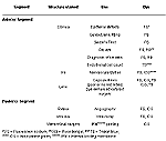
Various nontoxic ophthalmic dyes have been extensively used as diagnostic agents for the detection and management of different ocular disorders. Table 1 summarizes the use of various dyes in ophthalmology. Dyes such as fluorescein sodium and indocyanine green (ICG) have a long history of safety in humans.1 There have been an increasing number of reports of enhanced visualization by staining intraocular tissues during cataract surgery and vitreoretinal surgery.2-19 Staining of the ocular tissue by using the ophthalmic dyes makes visual differentiation and manipulation of tissues easier. Enhanced viewing of the ocular tissues can promote a surgeon's ability to evaluate clinical structural relationships and may help attain surgical objectives with fewer complications.2-19
Small-incision cataract surgery using phacoemulsification has evolved into one of the most successful surgical techniques in ophthalmology and the visual sciences. Many modifications such as continuous curvilinear capsulorrhexis (CCC),20,21 hydrodissection,22-25 hydrodelineation,26 and various maneuvers for nuclear emulsification and cortical cleanup have been added to it, increasing its safety and efficacy. Posterior capsulorrhexis, a technically challenging procedure, has also been recommended by several surgeons for delaying opacification of posterior capsule in pediatric cataracts and for managing the posterior capsule tears.27-31
We have extensively studied the use of nontoxic capsular dyes (fluorescein sodium, ICG, and trypan blue) to enhance visualization of intraocular tissues during various steps of the modern phacoemulsification procedure,7-9,12-14,32,33 which we will address here. We have divided this into three sections. In Section 1, we will discuss the use of capsular dye to stain the anterior capsule for CCC in mature white cataracts. In Section 2, we will discuss how capsular dye can enhance visualization as one learns the critical steps of phacoemulsification surgery: CCC, hydrodissection/hydrodelineation, nuclear emulsification, and cortical cleanup. Finally, in Section 3, we will address the use of posterior capsular dye staining in order to facilitate the challenging procedure of posterior capsulorrhexis.
Dye-Enhanced Anterior Capsulorrhexis
CCC offers unquestionable advantages over other capsulotomy techniques.20,21 Because of complications such as intraocular lens (IOL) asymmetrical fixation, decentration, or pea-podding of the IOL haptics associated with the envelope or the can-opener capsulotomy techniques, CCC is preferred in PE or planned extracapsular cataract extraction.20,21,34
Capsulorrhexis in Absence of Red Reflex?
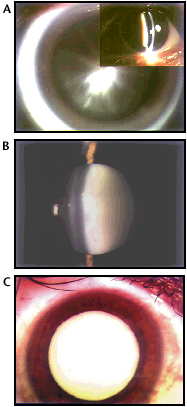
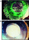
Mature white and hypermature cataracts are still commonly encountered, especially in the developing world (Slide 1 A, B, C).35
It is difficult to perform a CCC in the presence of mature cataracts because the red reflex, which is necessary to observe the actual tearing process,
is absent. With poor visibility, errant capsular tearing is very common and difficult to control, thus jeopardizing in-the-bag IOL implantation. The
accepted recommendations to aid CCC in such cases are dimming the operating room lights, increasing the operating microscope magnification
and coaxial illumination, and using high-molecular weight viscoelastics. The use of air,36 diathermy,37
endoilluminator,2 vitrectome, scissors,38 and the two-stage CCC approach33,39 have also been
suggested.
Capsular Dyes for Anterior Capsulorrhexis
Capsular dyes such as 2% fluorescein sodium, 0.5% ICG,5,7,10 and 0.1% trypan blue6,7,11,15 have been successfully
used to stain the anterior capsule for the purposes of performing CCC. Two main surgical techniques have been used for fluorescein sodium:
(a) staining from above, under an air bubble, and (b) intracameral subcapsular injection.3,4 Use of 0.001% to 0.01% gentian
violet solutions and also 0.05% to 0.25% crystal violet solutions have also been recently reported for staining the anterior capsule in animal models
(albino rats and rabbits, respectively).16,17 Gentian violet and crystal violet dyes are not preferred for human eyes due to adverse corneal
effects and possible endothelial cell toxicity.
Study Comparing Three Capsular Dyes and Two Surgical Techniques
We have evaluated, in an experimental closed-system surgery, anterior capsule staining for performing CCC in postmortem human eyes with
advanced/white cataracts, using three dyes.7 These are fluorescein sodium, ICG, and trypan blue; all have been clinically
advocated for use with this procedure. We also compared the two commonly used methods of staining under an air bubble and intracameral
subcapsular injection.7,40
A 2% fluorescein sodium solution was prepared by mixing 1 mL of 10% fluorescein for intravenous use (Alcon Ophthalmic, Fort Worth, Texas) with 4 mL of balanced salt solution (BSS). ICG solution (Akorn, Inc., Buffalo Grove, Ill) was prepared by dissolving 25 mg of ICG in 0.5 mL of an aqueous solvent (provided with the ICG), which was mixed in 4.5 mL of an irrigating solution (BSS plus, Alcon Ophthalmic, Fort Worth, Texas).5 To obtain 0.1% trypan blue, we mixed 1 mL of a 0.4 % solution (Life Technologies, Grand Island, NY) in 3 mL of BSS.
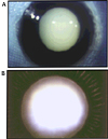
Randomly accessioned postmortem human eyes (n=12), received within 4 days of death in the Center for Research on Ocular Therapeutics and Biodevices from Eye Banks nationwide, were used in this study. We only used eyes presenting advanced/white cataracts (Slide 2 A, B). They were immersed in dextran solution for 30 minutes and were prepared according to the technique of Auffarth and colleagues.41 After the eye was fixed in the training head, a self-sealing corneoscleral tunnel incision approximately 3.2-mm wide was made. The iris was pulled out from its attachment to allow better visualization of the anterior capsule. Two different techniques were used for the capsular staining. Initially in six globes, air was carefully injected using a 27-gauge cannula and a 2.0-cc syringe. Then, the dye was injected over the anterior capsule (0.10 mL) within the air bubble (2 globes/dye). After a few seconds, the air bubble was replaced with sodium hyaluronate (Healon, Pharmacia), and CCC was then performed (Slide 3 A, B, C and Slide 3 D, E).
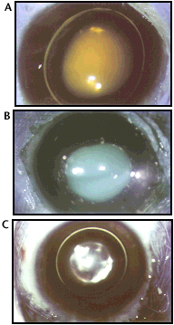
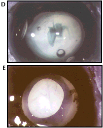
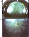
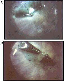
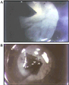
Alternatively, in the other six globes, we used the technique of intracameral subcapsular injection (Slide 4 A, B, Slide 4 C, D and Slide 5 A, B). After the aqueous was replaced with Healon, we carefully injected 0.05 mL to 0.10 mL of the dye beneath the anterior capsule (2 globes/dye) using a 30-gauge needle. A small leakage of dye from the subcapsular space was observed during this step. After the stained viscoelastic was replaced by clear Healon, the point of injection was used for beginning the CCC with Utrata’s forceps. Blue light enhancement was used during CCC for fluorescein sodium.
To compare the two techniques and the three dyes, the following two parameters were evaluated by two independent surgeons (SKP, LW):
1) ability to perform the staining technique (+ = difficult; ++ = intermediate; +++ = easy).
2) staining of the anterior capsule (+ = faint; ++ = intermediate; +++ = good).
Photographs were taken using a Topcon camera fitted to the operating microscope, with and without a blue filter. The Miyake-Apple posterior video/photographic technique42,43 was also used in another three globes to document any dye leakage into the vitreous cavity (1 globe/dye). We performed the staining of the anterior capsule under an air bubble using fluorescein sodium, ICG, and trypan blue in six globes (2 globes each). Alternatively, the same dyes were used for intracameral subcapsular injection in the other six globes (2 globes each).
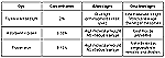
Table 2 shows the results of our evaluation of the two staining techniques (under an air bubble and intracameral subcapsular injection) and the three dyes (fluorescein sodium, ICG, and trypan blue). In all globes, CCC was completed uneventfully. Of these two techniques, the intracameral subcapsular injection provided a slightly better staining of the anterior capsule. The dye remained trapped in the subcapsular space after the injection, in contact with the posterior surface of the anterior capsule, allowing enough time to perform any maneuver. Among the three dyes used in this study, the staining provided by ICG, at the concentration used, was found to be slightly superior (Slide 4 A, B, Slide 4 C, D and Slide 5 A, B). It was notably easier to localize the ICG-stained posterior surface of the inverse anterior capsular flap while performing CCC, after the subcapsular injection of the dye (Slide 4 A, B and Slide 4 C, D). The Miyake-Apple posterior video/photographic technique demonstrated a leakage of fluorescein sodium into the vitreous after using both dye administration methods (under an air bubble and intracameral subcapsular injection). The intensity of this leakage increased progressively with time. Slide 6 A, B, C illustrates the progressive leakage of the fluorescein sodium into the vitreous cavity after intracameral subcapsular injection. No vitreous leakage was observed with ICG or trypan blue after using any of the two aforementioned techniques of anterior capsule staining.
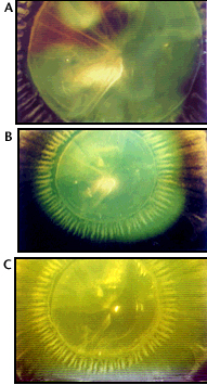
Table 3 and Table 4 summarize the advantages and disadvantages associated with the use of dyes (fluorescein sodium, ICG, and trypan blue) and the techniques of staining (under an air bubble and intracameral subcapsular injection).
Clinical Application and Guidelines for Surgeons
Staining of the anterior capsule with capsular dyes is a useful alternative for performing CCC in cases of advanced/white cataract. Fluorescein
sodium was the first dye advocated for this use.2-4 ICG and trypan blue were subsequently used for this purpose.5-7,10,11,15,18
ICG and trypan blue selectively stain dead corneal endothelial cells. Because the endothelial cells are alive in human cataract surgery, ICG and trypan
blue neither stain these cells nor obstruct the surgeon’s view. Because of its smaller molecular weight (376 d), fluorescein sodium can stain the cornea
and also migrate to the vitreous cavity. We were able to demonstrate the leakage of fluorescein sodium into the vitreous cavity using the Miyake-Apple
posterior video/photographic technique. In the study of Horiguchi and colleagues,5 fluorescein sodium could not be removed from the
vitreous cavity by an irrigation-aspiration system. The reconstituted ICG dye is only good for 10 hours. Because the bottle of ICG is expensive
(approximately US $90.00 for 25 mg ICG), it is better to schedule several patients with absent or poor red reflex on the same surgery day. Intraocular
solutions of trypan blue have not yet been approved by the Food and Drug Administration (FDA) and, therefore, are not currently available in the
United States; thus, the solution has to be prepared before surgery.
As it was proposed by Melles,6 trypan blue 0.1% solution (ready to be used) for capsular staining is commercialized
by DORC International (VN Zuidland, Holland) under the trade name of VisionBlue. Recently, a 0.1%
solution of trypan blue dye became commercially available from Dr. Agarwal's Pharma Ltd. (Chennai, India) under the trade name of Blurhex.
Trypan blue is less expensive than ICG, and, to the best of our knowledge, the cost of a 1-mL ampule of trypan blue (Blurhex) is approximately US $3.00,
compared to the US $90.00 cost of one ampule of 25 mg ICG powder.18
In some animal studies, when trypan blue dye was given intravenously or intraperitoneally (more frequent applications and at much higher concentrations), it induced neoplasms.44 For this reason, surgeons should probably avoid using this dye in pregnant women or children.
The surgeon should also be careful when using any capsular dyes in cataract surgery combined with implantation of hydrophilic acrylic lenses having a high water content (73.5%), as this can lead to permanent staining (discoloration) of the IOL by capsular dyes. This discoloration may also be associated with a decrease or alteration in the best-corrected visual acuity, eventually requiring IOL explantation/exchange. We have recently analyzed two Acqua hydrophilic acrylic lenses explanted secondary to bluish discoloration after use of trypan blue dye.45
The techniques originally reported for staining the anterior capsule using fluorescein sodium are staining from above, under an air bubble, as proposed by Nahra and Castilla in 1996 at the annual meeting of the American Academy of Ophthalmology46 and intracameral subcapsular injection of fluorescein sodium (staining from below) with blue-light enhancement.3,4 The first technique (staining under an air bubble) is currently used by most surgeons. One benefit is the staining of the peripheral anterior rim, which is otherwise difficult to visualize during the phacoemulsification procedure.6 However, air in the anterior chamber makes it unsteady. Any instrument entering the eye will cause some air to escape, with a rise of the lens-iris plane. A small amount of high-density viscoelastic placed near the incision can prevent the air bubble from escaping the anterior chamber, thus minimizing the risk of sudden collapse. Also, with this technique, there is a progressive dilution of the dye by the aqueous humor. This may be a possible explanation for the fainter staining observed with this technique in recent clinical reports.47 Most of the drawbacks of this technique can be avoided by careful use of a viscoelastic solution to seal the incision site. Akahoshi proposed the "soft shell stain technique" for performing a CCC in white cataract cases.48 A small amount of viscoelastic (Viscoat) was injected into the anterior chamber followed by high molecular weight viscoelastic material (Provisc) to fill up the chamber completely. The author then injected ICG solution on the lens surface with a bent G27 viso cannula. The anterior capsule was uniformly stained green and was easily visualized while the cornea remained unstained. According to the author, the soft shell stain technique is extremely useful for CCC of white cataracts.
Alternatively, the dye solution can be mixed with viscoelastic agents. Kayikicioglu and colleagues15 proposed a technique for limiting the contact of trypan blue by mixing the dye with a viscoelastic solution. These researchers mixed 0.4% trypan blue with 1% sodium hyaluronate in a 2-mL syringe. The dye, mixed in a viscoelastic solution, is injected onto the anterior lens capsule, which covers the anterior capsule without coming in contact with the corneal endothelium. Trypan blue mixed with sodium hyaluronate greatly increases the visibility of the anterior lens capsule without staining the adjacent tissues. There is a potential risk of corneal decompensation after intraocular use of self-mixed solution; however, these authors used this technique without significant surgical and postoperative adverse effects.
Intracameral subcapsular injection is another, but less commonly used, technique for anterior capsule staining. It has the advantage of trapping the dye in the central and midperipheral subcapsular space. It gives sufficient time for the surgeon to perform any maneuver until the CCC releases it. Meanwhile, the dye remains in contact with the posterior surface of the anterior capsule. This may be a possible explanation for the better staining observed in our laboratory study on postmortem human eyes.7 The capsule and cortex are both stained by the dye used, but they can be clearly distinguished from the feathery appearance of the cortex and the smooth staining of the capsule. The CCC is fairly easy to perform by grasping the injection hole. This technique was originally proposed for fluorescein with blue-light enhancement, but we also used it with ICG and trypan blue. When the capsular flap is inverted, the stained posterior surface of the anterior capsule enhances visualization and thus facilitates tearing during CCC. In our study, this was more obvious with the intracameral subcapsular injection of ICG.7 This can be performed without the need for any special type of illumination, such as a cobalt blue-filter. We would like to emphasize that there is the risk of anterior capsule tear formation after subcapsular injection of dye. However, we did not observe this complication (anterior capsule tear) in any postmortem human eyes used in our laboratory study.7 We would certainly not recommend the intracameral subcapsular injection technique for performing CCC in intumescent and hypermature cataracts owing to the high intralenticular pressure and fragile anterior lens capsule that may easily lead to radial tear formation.
Horiguchi and colleagues5 reported the technique of staining the anterior capsule using a 2% solution of ICG in patients with mature cataracts. They compared the results of phacoemulsification and IOL implantation in two groups of 10 eyes. In the first group, the anterior capsule was stained with ICG before CCC, and in the second, no dye was used. There was no statistically significant difference reported in their study between both groups concerning specular-microscopy endothelial cell counting and laser flare-cell photometry; thus, the staining procedure was considered to be safe.
Chang has reported his clinical experience with ICG and trypan blue for anterior capsule staining in mature white or brunescent cataracts. The author reported his clinical experience in two consecutive, non-randomized series of mature or brunescent cataracts. Using the technique of dye injection under an air bubble, ICG dye was used in the first series, and trypan blue in the subsequent series. Both dyes provided consistently excellent visualization and clinical results without any adverse effects. However, trypan blue created a more intense and persistent staining and provided superior visualization when compared with ICG, according to this first clinical study.49,50
We would like to provide some recommendations and guidelines for ophthalmic surgeons regarding suitable capsular dyes and the anterior capsule staining technique. These are based on our experience in postmortem human eyes, as well as published clinical reports from several other surgeons. Both ICG and trypan blue are currently preferred over fluorescein sodium dye, due to better staining of the anterior capsule and the absence of vitreous leakage (due to high molecular weight). Both of these dyes provide excellent visualization of the anterior capsule flap during CCC, without causing any toxic effects on the corneal endothelium. Trypan blue has the advantage of being less costly when compared to the cost of ICG. However, trypan blue should be avoided in fertile/pregnant women and in children due to the possible teratogenic and/or mutagenic effects observed in animal studies when using this dye. Further studies may be helpful to determine the least available concentration of the trypan blue dye that can be used to stain the anterior lens capsule in order to perform the CCC. ICG remains a valuable alternative for these special patients (children and pregnant women). Staining under the air bubble technique is safer and is therefore recommended for intumescent and hypermature cataract patients presenting with high intralenticular pressure and a fragile anterior lens capsule. Viscoelastic solutions can be used to visco-seal the incision site in order to avoid escape of the air bubble and to minimize any anterior chamber fluctuations. Alternatively, mixing the dye with a viscoelastic solution may also be used for better anterior capsule staining and for limiting the contact with adjacent ocular tissues.
Dye-Enhanced Phacoemulsification
For the beginning cataract surgeon, there are benefits to practicing the phacoemulsification procedure in a wet laboratory setting. This can shorten the learning curve and improve safety before operating on actual patients.51-54 Human eye bank or animal eyes are commonly used for this purpose. The surgeon must be familiar with the critical steps of the phacoemulsification procedure. Each step is therefore to be learned independently and carefully in order to achieve a successful outcome and reduce complications. This is even more important when dealing with advanced/white cataracts in a clinical setting. The absence of a red reflex in such cases renders CCC as well as nucleus sculpting maneuvers difficult, if not impossible.55
In an earlier section, we discussed staining of the anterior capsule for CCC in advanced cataracts, comparing three dyes (fluorescein sodium, ICG, and trypan blue dyes) and two injection techniques in postmortem human eyes. All dyes provided satisfactory staining of the anterior capsule for CCC. However, fluorescein sodium progressively leaked into the vitreous.7 In this section, we report our experience with the application of 0.5% ICG and 0.1% trypan blue to obtain complete hydrodissection/delineation, to stain the nuclear substance during nuclear emulsification, and to stain the capsular bag during the cortical cleanup. Application of the capsular dye for performing phacoemulsification is termed dye-enhanced phacoemulsification, and this section will focus on the use of dye to enhance visualization for learning and performing critical steps of the phacoemulsification procedure.
Study of Dye-Enhanced Phacoemulsification: Surgical Technique
Randomly accessioned postmortem human eyes (n = 16) obtained within 4 days of death from eye banks nationwide were used in our study.8
They were prepared according to the Miyake-Apple posterior video technique.42,43 Two independent surgeons (LW, SKP) evaluated the use of 0.5% ICG and 0.1% trypan blue to perform the critical steps of phacoemulsification in eight eyes (two eyes/dye/surgeon). Dye solutions were prepared as previously described. In eight other eyes used as controls, the phacoemulsification procedures were performed without the use of dye. After the cornea and iris were removed, a CCC (4.5 to 5.5 mm in diameter) was initiated using a 26-gauge needle cystotome and was completed using Utrata’s forceps. A complete cortical cleaning hydrodissection was performed by injecting 2 to 3 cc of the dye solution (0.5% ICG or 0.1% trypan blue) between the lens capsule and the cortex with a 27-gauge cannula. This was followed by hydrodelineation: placement of a 27-gauge cannula deep into the nucleus and injection of the dye solution created the colored fluid-wave marking the separation of the nucleus and the epinucleus. Balanced salt solution (BSS) was used to perform hydrodissection/delineation in the control group. Nuclear emulsification (Alcon Legacy 20,000, Alcon Surgical, Fort Worth, TX) was performed using the divide-and-conquer nucleofractis technique.55 One to 2 microdrops of the dye solution were instilled into the capsular bag, and cortical cleanup was performed using the irrigation and aspiration system.
The enhancement of visualization while performing each step of the surgery using the dyes was evaluated by the two independent surgeons. They particularly noted:
- if the use of a colored solution helped in visualizing the fluid waves and the plane of cleavage during hydrodissection/delineation
- if the staining of the nucleus substance helped in appreciating the depth of the phaco tip and its position in relation to the posterior capsule during the nuclear emulsification
- if the staining of the inner surface of the capsular bag helped in identifying residual cortical material during the cortical cleanup procedure using the irrigation-aspiration system
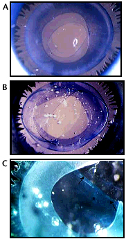
Our experimental study suggested that both dyes (0.5% ICG and 0.1% trypan blue) successfully enhanced visualization while performing critical steps of the phacoemulsification procedure, when compared to the control group.8 During hydrodissection/hydrodelineation, the use of dye helped to visualize the formation of a complete cleavage between the capsule and cortex, as well as between the nucleus-epinucleus complex (Slide 7 A, B). Incomplete cleavage could be easily identified by using a colored solution and could be promptly completed by re-injection in the appropriate quadrant.
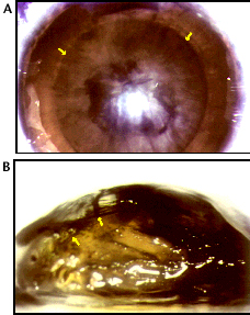
During the nuclear emulsification procedure, the use of dye helped in appreciating the position of the phaco tip and its relation with the posterior capsule, thus increasing the safety of the procedure (Slide 8 A, B).
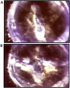
For the complete cleaning of the capsular bag, the use of dye provided better visualization of the residual cortical material during the irrigation-aspiration procedure (Slide 9 A, B). It was easy to differentiate the feathery, irregular staining of the cortex from the smooth staining of the capsule.
Learning Critical Steps of Phacoemulsification
The critical steps of the phacoemulsification procedure are CCC, hydrodissection, hydrodelineation, nuclear emulsification maneuvers, and cortical
cleanup. In a series of 7,169 patients undergoing phacoemulsification, Gimbel reported 36 preoperative posterior capsule tears.56
Of these, 19 (53%) occurred during the irrigation/aspiration step, and 13 (36%) occurred during the phacoemulsification itself. Five percent and 6%
of the posterior capsule tears occurred during the IOL implantation and the hydrodissection, respectively. Visualization while performing each step
can be enhanced by the use of different dyes, thus possibly enhancing safety.
CCC
We have addressed the techniques of anterior capsule staining for anterior capsulorrhexis using fluorescein sodium, ICG, and trypan blue
dyes. In this section, we used only ICG and trypan blue, because of the vitreous leakage associated with fluorescein sodium. In addition to the
staining of the anterior capsule, ICG and trypan blue can also be successfully used to learn other critical steps of the phacoemulsification procedure
in a laboratory setting. We believe that the use of dye would not only be useful during the anterior capsule management in white cataracts, but also
for learning and practicing the other critical steps of phacoemulsification procedure as discussed here.
Hydrodissection/Hydrodelineation
Hydrodissection is an important step of small-incision cataract surgery using phacoemulsification. The use of this procedure was first reported by
Faust.22 Fine24 added the concept of cortical cleavage hydrodissection to separate the superficial cortex from the
lens capsule. The use of dye (instead of BSS) helps in the localization of the complete (360°) cortical cleavage plane, separating the equatorial and
posterior capsule from the cortex. It is much easier to visualize a dye-colored fluid wave of hydrodissection. Therefore, it helps in achieving complete
separation between capsule and cortex. Hydrodelineation,26 associated with the formation of a golden ring, is sometimes difficult to
notice. With the injection of a capsular dye solution, however, the surgeon can successfully visualize the demarcation between the nucleus and the
epinucleus (Slide 7 A, B). In this situation, an incomplete hydrodissection/delineation can be easily identified and completed by injecting more stained
fluid in that particular quadrant, if needed. After achieving complete hydrodissection and hydrodelineation, it is easier to perform nuclear emulsification
with less ultrasound power and time, decreasing the need for cortical cleanup and the risk of posterior capsule tears. Recent studies from our
laboratory suggest that hydrodissection-enhanced cortical cleanup is an unidentified important factor for delaying the onset of posterior capsule
opacification.57
Nuclear Emulsification
A number of different techniques have been used by surgeons for nuclear emulsification of hard and soft cataracts. While performing nuclear
emulsification maneuvers, visualization of the depth at which the phaco tip is sculpting is crucial. Its significance cannot be overemphasized for
preventing complications like unnoticed posterior capsular tears, vitreous loss, and dislocation of the nucleus into the vitreous cavity. When a
good red reflex is present, the surgeon can rely on an increasingly brighter-red reflex to gauge proximity to the posterior capsule while sculpting
the nucleus. However, the absence of a red reflex (e.g., in advanced, white, mature, or hypermature cataracts) complicates nucleus emulsification
because it is difficult to judge the depth of the phaco tip during sculpting.58 The staining of the nucleus (lens substance) helps in
visualizing the position of the phaco tip and its relation with the posterior capsule, thus enhancing the safety margin of the procedure.
Cortical Cleanup
Studies have shown that at least half of the cases of capsular tears and vitreous loss occur at the time of cortical cleanup.56
Staining of the capsular bag enhances its visualization, and the surgeon can distinguish feathery, irregular staining of residual cortex from smooth
staining of the anterior, equatorial, and posterior capsule. Thus, the staining facilitates the cleaning of residual cortical matter from the capsular bag.
Posterior capsule staining can also be very useful to learn and perform the posterior continuous curvilinear capsulorrhexis (PCCC) procedure, details
of which will be provided in the next section.
Possible Clinical Application and Future Trials
Our laboratory study provides evidence that both dyes (ICG and trypan blue) can be used in the clinical setting of living human eye operations to
achieve a complete hydrodissection/delineation.8 They can also be used to visualize the depth of the phaco tip during sculpting
and its relation to the posterior capsule, especially in patients with a poor or absent red reflex (advanced/white cataracts). It is easier to differentiate
the cortical matter from the anterior or posterior capsules after the staining of the capsular bag. This would be helpful in achieving a complete cortical
cleanup during the irrigation/aspiration steps, with lower incidence of posterior capsule tear.
In summary, the use of dyes (ICG and trypan blue) during small-incision cataract surgery using phacoemulsification facilitated complete hydrodissection and hydrodelineation, nuclear emulsification maneuvers, and a complete cortical cleanup. We believe that, in addition to practicing an anterior capsulorrhexis, these dyes can also be used to enhance visualization for learning other important steps of the phacoemulsification procedure in a wet laboratory setting.8 This study also suggests the need for future clinical trials using these dyes in a clinical setting of living human eye operations.
Dye-Enhanced Posterior Capsulorrhexis
PCCC is a posterior continuous central capsulotomy technique described by Gimbel and Blumenthal and colleagues in 1990.27,28 PCCC is recommended for converting an irregular tear of the posterior capsule into a circumscribed cut not extending to the equator.27,29 It can also be used for the removal of posterior capsular plaques in posterior subcapsular or polar cataracts.59
Recently, the use of PCCC combined with optic capture of an IOL30,31 and/or anterior vitrectomy60-63 successfully evolved for delaying the development of posterior capsule opacification (PCO) or secondary membrane formation in pediatric cases. Primary posterior capsulotomy, in the form of PCCC, is especially important in younger children for maintaining a long-term clear visual axis in order to prevent the development of amblyopia.64 Besides children, some surgeons have considered the controversial concept of PCCC in adults as an alternative to Nd:YAG laser capsulotomy for PCO.65,66
Learning to perform a PCCC is technically challenging due to the thin and transparent nature of the posterior capsule. Further, it should not be attempted if visibility is mediocre and/or vitreous pressure is assessed to be high.
Study of Dye-Enhanced Posterior Capsulorrhexis
We conducted a study in human eyes obtained postmortem to assess capsular dye-enhanced PCCC.9
Randomly accessioned postmortem human eyes (n = 12) obtained within 4 days of death from eye banks nationwide were used in this study. The eyes were prepared according to the Miyake-Apple posterior video technique.42,43 They were sectioned at the equator, and the anterior segment was mounted on a glass slide to provide a posterior perspective of this portion of the eye. After the cornea and iris were removed, a capsulorrhexis 5.0 to 5.5 mm in diameter was initiated using a 26-gauge needle and completed using a Utrata’s forceps. A complete cortical-cleavage hydrodissection was performed by injecting balanced salt solution (BSS, Alcon Ophthalmic, Fort Worth, TX) between the lens capsule and the cortex with a 27-gauge cannula. This was followed by careful hydroexpression of the nucleus, avoiding damage to the posterior capsule. Cortical cleanup was performed using an irrigation/aspiration system.
Two independent surgeons (SKP, LW) evaluated the use of dye to enhance visualization of the posterior capsule during PCCC (four eyes/surgeon). Both surgeons were inexperienced with the PCCC procedure and performed it for the first time in their professional careers. PCCC was also performed in four other eyes (two eyes/surgeon) without the use of dye. The posterior capsule was stained by instilling 1 microdrop of the dye solution into the capsular bag. We used 0.5% ICG and 0.1% trypan blue solutions (four eyes/dye). The dye solutions were prepared as described by us earlier. After waiting 1 to 2 minutes, the excessive dye was washed out. The capsular bag was filled with a viscoelastic agent (Healon, Pharmacia Inc., Peapack, NJ), and PCCC was initiated by using a 26-gauge needle cystotome and was completed using a Utrata’s forceps. Optic capture of a posterior chamber intraocular lens (PC-IOL), as well as an anterior vitrectomy, was also practiced.
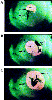
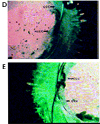
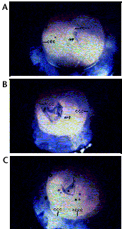
Our laboratory study in eight postmortem human eyes confirmed that the posterior capsule can be successfully stained using ICG and trypan blue (Slide 10 A, B, C, D, E and Slide 11 A, B, C, D, E). For both surgeons, it was much easier to initiate and complete PCCC successfully after staining of the posterior capsule, when compared to the control (non-stained) eyes. PCCC was completed fairly easily in all globes, due to better visualization of the stained posterior capsule flap (PCF) against the transparent (non-stained) anterior hyaloid phase (AHP) of the vitreous (Slide 10 A, B, C and Slide 11 A, B, C). It was simpler to perform optic capture of PC-IOLs after staining the posterior capsule (Slide 10 D, E and Slide 11 D, E). Both dyes used in this study provided satisfactory visualization of the capsule during PCCC.
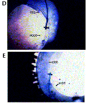
Clinical Application and Future Trials
PCCC is currently attracting more interest as a means of PCO prevention in children. Additionally, this procedure is also important in the
management of posterior capsule tears of congenital, traumatic, or surgical origin and the pealing of plaques associated with posterior polar and
posterior subcapsular cataracts.59 As mentioned earlier, PCCC is technically challenging due to the thin, transparent, and elastic
nature of the posterior capsule. Attempting PCCC in the presence of poor visibility associated with positive vitreous pressure is difficult and may
cause an inadvertent radial tear extending toward the equator. Cauwenberge, Rakic, and Galand67 recently reported on the etiology,
management, and outcome of complicated posterior capsulorrhexis. In a 1-year retrospective analysis of 650 patients, they identified 32 (5%)
cases of complicated PCCC. According to them, the most frequent problem was the performance of a central capsulorrhexis within the optimum
size (<5 mm in diameter). This was not possible in 14 (44%) cases in their series. In 12 (37%) cases, a PCCC was carried out with great difficulty
because of either insufficient visibility during the procedure or an anatomically changed capsule (floppy or fibrotic in its center). Vitreous loss occurred
in six (19%) cases during the PCCC procedure. All the aforementioned complications were reported by these authors despite their 4-year experience
with the PCCC procedure, performed on more than 1,300 patients.
Posterior capsulorrhexis, which is considered technically difficult and challenging, became easier to learn and perform after staining the posterior capsule with ICG or trypan blue in the laboratory setting. Staining enhances visualization of the posterior capsule flap, which can be easily recognized against the transparent anterior hyaloid phase of the vitreous.
Dye-Enhanced Pediatric Cataract Surgery
We have also evaluated the use of two capsular dyes, 0.5% ICG and 0.1% trypan blue, for staining the posterior capsule, while performing PCCC in
pediatric eyes obtained postmortem (Slide 12 A, B, C).14 Pediatric eyes are associated with thin sclera and positive vitreous pressure,
making PCCC especially difficult.61,62,64 Gimbel’s technique of PCCC with IOL optic capture might be facilitated by dye staining
of the posterior capsule.30,31 Depending on the type of dye used, vitreous loss can be easily identified by the formation of colored
localized clumps.
In brief, results of our laboratory study in postmortem human eyes revealed that posterior capsule staining using ICG or trypan blue is very helpful when performing the PCCC procedure in adults and children. Clinical trials using these dyes to stain the posterior capsule for PCCC are needed. This technique would be particularly valuable for the beginning surgeon, or in the presence of poor visibility.
Summary and Conclusions
The use of non-toxic capsular dyes to enhance visualization during the various steps of cataract surgery has been summarized in Table 5. Based on our experience of dye-enhanced cataract surgery, we confirm that capsule staining using various dyes in cataractous eyes with poor or no red reflex helps create an intact anterior capsulorrhexis.7 Several laboratory and clinical studies suggested that both ICG and trypan blue are safe and provide excellent visualization of the anterior capsule flap during CCC.5-7,10,12,13,50,68 The trypan blue dye has the advantage of being less expensive than ICG. However, trypan blue dye should not be used in fertile/pregnant women or in children due to the possibility of a teratogenic and/or mutagenic effect. ICG remains a valuable alternative for these special patients. Anterior capsule staining under the air bubble technique provides good visualization without the possibility of an anterior capsule tear formation; thus, it is suitable in intumescent and hypermature cataract patients. In addition, our laboratory studies demonstrate that the capsular dyes can be successfully used to enhance visualization while learning the other critical steps of phacoemulsification.8 Also, the technically more challenging procedure of posterior capsulorrhexis can be performed with more facility.9
References
- Hogan RN, Zimmerman CF. Sodium fluorescein and other tissue dyes. In: Zimmerman TJ, et al. Textbook of Ocular Pharmacology. Philadelphia, PA: Lippincott-Raven; 1997:849-863.
- Mansour AM. Anterior capsulorhexis in hypermature cataracts (letter). J Cataract Refract Surg. 1993;19:116-117.
- Hoffer KJ, McFarland JE. Intracameral subcapsular fluorescein staining for improved visualization during capsulorhexis in mature cataracts (letter). J Cataract Refract Surg. 1993;19:566.
- Fritz WL. Fluorescein blue light assisted capsulorhexis for mature or hypermature cataract. J Cataract Refract Surg. 1998;24:19-20.
- Horiguchi M, Miyake K, Ohta I, Ito Y. Staining of the lens capsule for circular continuous capsulorhexis in eyes with white cataract. Arch Ophthalmol. 1998;116:535-537.
- Melles GRJ, Waard PWT, Pameyer JH, Beekhuis WH. Trypan blue capsule staining in cataract surgery. J Cataract Refract Surg. 1999;24:7-9.
- Pandey SK, Werner L, Escobar-Gomez M, Roig-Melo EA, Apple DJ. Dye-enhanced cataract surgery. Part 1: anterior capsule staining for capsulorhexis in advanced/white cataract. J Cataract Refract Surg. 2000;26:1052-1059.
- Werner L, Pandey SK, Escobar-Gomez M, Hoddinott DSM, Apple DJ. Dye-enhanced cataract surgery. Part 2: learning critical steps of phacoemulsification. J Cataract Refract Surg. 2000;26:1060-1065.
- Pandey SK, Werner L, Escobar-Gomez M, Werner LP, Apple DJ. Dye-enhanced cataract surgery. Part 3: Posterior capsule staining to learn posterior continuous curvilinear capsulorhexis. J Cataract Refract Surg. 2000;26:1066-1071.
- Newsom TH, Oetting TN. Indocyanine green staining in traumatic cataract. J Cataract Refract Surg. 2000;26:1691-1693.
- Nodarian M, Feys J, Sultan G, Salvanet-Bouccara A. Capsulorhexis staining by trypan blue in mature cataract surgery. J Fr Ophtalmol. 2001;24:274-276.
- Pandey SK, Werner L, Apple DJ. Staining the anterior capsule (letter). J Cataract Refract Surg. 2001;27:647-648.
- Pandey SK, Werner L, Apple DJ, et al. Capsular dye-enhanced cataract surgery. In: Nema HV, ed. Recent Advances in Ophthalmology, Vol. 5. New Delhi, India: Jaypee Brothers; (in press).
- Pandey SK, Werner L, Apple DJ, et al. Dye-enhanced pediatric cataract surgery. J Pediatr Ophthalmol Strabismus. (submitted).
- Kayikicioglu O, Erakgun T, Guler C. Trypan blue mixed with sodium hyaluronate for capsulorhexis. J Cataract Refract Surg. 2001;27:970.
- Unlu K, Askunger A, Soker S, et al. Gentian violet solution for staining the anterior capsule. J Cataract Refract Surg. 2000;26:1228-1232.
- Gamal Eldin SA, el Mehelmy EM, el Shazli Mostafa YM. Experimental staining of the anterior lens capsule in albino rabbits. J Cataract Refract Surg. 1999;25:1289-1294.
- Agarwal A, Agarwal A, Agarwal S. Trypan blue in the management of mature cataracts. In: Agarwal S, Agarwal A, Sachdev MS, Mehta KR, Fine IH, Agarwal A, eds. Phacoemulsification, Laser Cataract Surgery and Foldable IOLs. New Delhi, India: Jaypee Brothers; 2000:618-623.
- Burke SE, DaMata AP, Snyder ME, et al. Indocyanine green-assisted peeling of the internal limiting membrane. Ophthalmology. 2000;107:2010-2014.
- Gimbel HV, Neuhann T. Development, advantage, and methods of the continuous circular capsulorhexis technique. J Cataract Refract Surg. 1990;16:31-37.
- Assia EI, Apple DJ, Barden A, et al. An experimental study comparing various anterior capsulotomy techniques. Arch Ophthalmol. 1991;109:642-647.
- Faust KJ. Hydrodissection of soft nuclei. Am Intraocular Implant Soc J. 1984;10:75-77.
- Anis AY. Origin of hydrodissection (reply—letter to the editor). J Cataract Refract Surg. 1995;21:6.
- Fine IH. Cortical cleaving hydrodissection. J Cataract Refract Surg. 1992;18:508-512.
- Koch DD, Liu JF. Multilamellar hydrodissection in phacoemulsification and planned extracapsular surgery. J Cataract Refract Surg. 1990;16:559-562.
- Gimbel HV. Hydrodissection and hydrodelineation. Int Ophthalmol Clin. 1994;34:73-90.
- Gimbel HV. Posterior capsule tears using phacoemulsification: causes, prevention and management. Eur J Implant Refract Surg. 1990;2:63-69.
- Blumenthal M, Assia E, Neumann D. The round capsulorhexis capsulotomy and the rationale for 11.0 mm diameter IOL. Eur J Implant Refract Surg. 1990;2:15-19.
- Castaneda VE, Legler UF, Tsai JC, et al. Posterior continuous capsulorhexis. An experimental study with clinical applications. Ophthalmology. 1992;2:63-69.
- Gimbel HV, DeBroff BM. Posterior capsulorhexis with optic capture: maintaining a clear visual axis after pediatric cataract surgery. J Cataract Refract Surg. 1994;20:658-664.
- Gimbel HV. Posterior capsulorhexis with optic capture in pediatric cataract and intraocular lens surgery. Ophthalmology. 1996;103:1871-1875.
- Pandey SK, Werner L, Escobar-Gomez M, Apple DJ. Anterior capsule staining in advanced cataracts: A laboratory study using postmortem human eyes. Presented at the meeting of the American Academy of Ophthalmology, Orlando, Fla., October 1999.
- Pandey SK, Werner L, Apple DJ, et al. Dye-enhanced cataract surgery in human eyes obtained postmortem: a laboratory study to learn critical steps of phacoemulsification. Presented at the XVII Congress of the European Society of Cataract and Refractive Surgery, Vienna, Austria, September 1999.
- Pande M. Continuous curvilinear (circular) capsulorhexis and planned extracapsular cataract extraction: are they compatible? Br J Ophthalmol. 1993;77:152-157.
- Vasavada AR, Singh R, Desai J. Phacoemulsification of white cataracts. J Cataract Refract Surg. 1998;24:270-277.
- Brusini P. Capsulorhexis in mature cataracts: Why not? Doc Ophthalmol. 1992;81:281-284.
- Hausmann N, Richard G. Investigations on diathermy for anterior capsulotomy. Invest Ophthalmol Vis Sci. 1991;32:2155-2159.
- Vajpeyee RB, Angra SK, Honavar SG, et al. Capsulotomy for phacoemulsification in hypermature cataracts. J Cataract Refract Surg. 1995;21:612-615.
- Gimbel HV. Two stage capsulorhexis for endocapsular phacoemulsification. J Cataract Refract Surg. 1990;16:246-249.
- Pandey SK, Werner L, Apple DJ. ICG staining reduces your risk, compare three dyes. In: Clinical Update: Cataract. EyeNet. 2000;4:25-26.
- Auffarth GU, Wesendahl TA, Solomon K, et al. A modified preparation technique for closed system ocular surgery of human eyes obtained postmortem: an improved research and teaching tool. Ophthalmology. 1996;103:977-988.
- Miyake K, Miyake C. Intraoperative posterior chamber lens haptic fixation in the human cadaver eye. Ophthalmic Surg. 1985;16:230-236.
- Apple DJ, Lim ES, Morgan RC, et al. Preparation and study of human eyes obtained postmortem with the Miyake posterior photographic technique. Ophthalmology. 1990;97:810-816.
- Melles GRJ. Trypan blue dye helps to visualize capsulorhexis. Cataract and Refractive Surgery Eurotimes. 1999;May-June.
- Werner L, Apple DJ, Crema A, et al. Permanent bluish discoloration of a hydrophilic intraocular lenses caused by intraoperative use of trypan blue. J Cataract Refract Surg. (in press).
- Nahra D, Castilla M. Capsulorhexis in no view cataract: staining of the anterior capsule with 2% fluorescein. Presented at the annual meeting of the American Academy of Ophthalmology, Chicago, October 1996.
- Steinert RF. ICG dye aids in visualization of the anterior capsule. Ophthalmology Times. May 15, 1999.
- Akahoshi T. Soft shell stain technique for white cataract. Presented at the ASCRS symposium on Cataract, IOL, and Refractive Surgery, Boston, May 2000.
- Chang DF. Capsule staining and mature cataracts: a comparison of indocyanine green and trypan blue dyes. Br J Ophthalmol (video report). July 2000.
- Chang DF. Compare two dyes. EyeNet. 2000;4:22.
- Pandey SK, Werner L, Escobar-Gomez M, et al. Creating cataracts of varying hardness to practice extracapsular cataract extraction and phacoemulsification. J Cataract Refract Surg. 2000;26:322-329.
- Pandey SK, Werner L, Vasavada AR, Apple DJ. Induction of cataracts of varying degrees of hardness in human eyes obtained postmortem for cataract surgeon training. Am J Ophthalmol. 2000;129:557-558.
- Synder RW, Donnenfeld ED. Teaching phacoemulsification to residents and physicians in transition. Int Ophthalmol Clin. 1994;34:191-199.
- Synder RW, Allinson RW, Fante RG, et al. Learning phacoemulsification surgery. Ophthalmology. 1992;99:448-453.
- Gimbel HV. Divide and conquer nucleofractis phacoemulsification and variations. J Cataract Refract Surg. 1991;17:281-291.
- Gimbel HV. Posterior capsule tears using phacoemulsification: causes, prevention and management. Eur J Implant Refract Surg. 1990;2:63-69.
- Peng Q, Apple DJ, Visessook N, et al. Surgical prevention of posterior capsule opacification. Part II. Enhancement of cortical clean up by focusing on hydrodissection. J Cataract Refract Surg. 2000;26:188-197.
- Chang D. Solutions offered for mature white cataracts. Ocular Surgery News. 1999;6:8.
- Vasavada A, Singh R. Phacoemulsification in eyes with posterior polar cataract. J Cataract Refract Surg. 1999;25:238-245.
- Basti S, Ravishankar U, Gupta S. Results of a prospective evaluation of management of pediatric cataracts. Ophthalmology. 1996;103:713-720.
- Pandey SK, Wilson ME, Trivedi RH, et al. Pediatric cataract surgery and intraocular lens implantation: current techniques, complications and management. Int Ophthalmol Clin. 2001;41:175-196.
- Wilson ME, Pandey SK, Werner L, Ram J, Apple DJ. Pediatric cataract surgery: current techniques, complications and management. In: Agarwal S, Agarwal A, Sachdev MS, Mehta KR, Fine H, Agarwal A, eds. Phacoemulsification, Laser Cataract Surgery and Foldable IOLs. New Delhi, India: Jaypee Brothers; 2000:369-388.
- Ram J, Pandey SK. Infantile cataract surgery: Current techniques, complications and their management. In: Dutta LC, ed. Modern Ophthalmology. New Delhi, India, Jaypee Brothers; 2000:378-384.
- Vasavada A, Desai J. Primary posterior capsulorhexis with and without anterior vitrectomy in congenital cataracts. J Cataract Refract Surg. 1997;23:645-651.
- Galand A, Cauwenberge VF, Moosavi J. Posterior capsulorhexis in adult eyes with intact and clear capsules. J Cataract Refract Surg. 1996;22:458-461.
- Galand A. Primary posterior capsulorhexis in adults. In: Nema HV, Nema N, eds. Recent Advances in Ophthalmology 4. New Delhi, India: Jaypee Brothers; 1998:157-161.
- Cauwenberge VF, Rakic JM, Galand A. Complicated posterior capsulorhexis: aetiology, management, and outcome. Br J Ophthalmol. 1997;81:195-198.
- Sharma N, Pangtey MS, Dada VK. Experience with indocyanine green dye (letter). J Cataract Refract Surg. 2001;27:1342.
*This was prepared with modification of our original published work on Dye-Enhanced Cataract Surgery (Part 1, 2, and 3) in the Journal of Cataract and Refractive Surgery, July 2000 issue.