Reprinted with permission from Masket S. Cataract surgery complicated by the miotic pupil. In: Buratto L, Osher RH, Masket S, eds. Cataract Surgery in Complicated Cases. 2000:131-137.
Introduction
Among the operative challenges faced by the cataract surgeon, the miotic pupil is the most common obstacle. The miotic pupil is also a condition that is associated with the greatest likelihood for intraoperative complications. A study of 1000 consecutive extracapsular cataract extractions demonstrated that the poorly dilating pupil was the most significant factor associated with capsule rupture and vitreous loss.1 Because patients with small pupils undergoing cataract surgery are at significant risk, surgeons must develop a plan for handling these cases while maintaining an acceptable rate of complications.
In the past, surgeons have altered and often misshaped the iris intraoperatively to eliminate the obstacles of the miotic pupil. A typical result of early cataract removal procedures, as noted in Slide 1, was accompanied by a significant change in the appearance of the iris. The postoperative aesthetic consequences of such surgery are potentially troublesome to patients because patients often judge the quality of surgery by the appearance of the postoperative eye in addition to the clarity of vision.
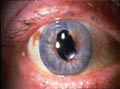
Surgical alteration of the pupil can have significant bearing on pupillary function with consequential iatrogenic-induced glare dysfunction. In a 1992 investigation, I reported that an enlarged pupil can be responsible for postoperative glare disability in eyes that were anatomically normal except for having pseudophakia.2 Because cataract surgery is often performed for glare symptoms, it is imperative that surgical methods do not induce other causes of postoperative glare. Therefore, a surgeon considering the preoperative patient with a miotic pupil is challenged by the increased likelihood for intraoperative complications, the need to maintain the aesthetics of the iris, and the mandate to respect the function of the pupil. The approach to the patient with a poorly dilating pupil may vary according to the cause of the presurgical miosis. The cause may vary from simple dilator atrophy noted commonly in elderly patients to extensive posterior synechiae from ocular inflammatory disease.
Additionally, long-term use of parasympathomimetic agents for glaucoma management will induce miosis with or without pupillary synechiae. Table 1 represents a partial list of the various causes for the poorly dilating pupil. The surgical approach at the time of cataract removal should vary according to the cause of the small pupil.
Table 1. Causes of Miotic Pupil
- Age-related dilator atrophy
- Diabetes
- Chronic use of miotics
- Idiopathic complications
- Intraoperative trauma
- Marfan syndrome
- Prior surgery (e.g., postvitrectomy, trabeculectomy)
- Pseudoexfoliation
- Synechiae
- Uveitis
Preoperative Therapy
Management of the miotic pupil begins with a careful preoperative evaluation. If the patient's history reveals prolonged use of miotics for glaucoma management, these agents should be stopped at an appropriate safe interval before surgery. If necessary, intraocular pressure (IOP) can be managed with alternative drugs, unless contraindicated. Physical examination will demonstrate the ability of the pupil to dilate in response to trial pharmacological stimulation. Inflammatory pupillary membranes, posterior synechiae, and pseudoexfoliation (Slide 2) will be evidenced and surgical plans for the pupil can be considered. In certain cases, nonspecific resistance to dilation will be observed.
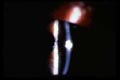
In preparation for surgery, pupil dilation is accomplished with three topical agents, a cycloplegic, a mydriatic, and a nonsteroidal anti-inflammatory drug (NSAID).
Cycloplegics
The following medications are commonly used clycloplegics:
- Cyclopentolate hydrochloride 1% provides both pupil dilation and cycloplegia and is the most commonly used cycloplegic agent. It is long acting; the pupil may stay dilated for as long as 36 hours following surgery.
- Tropicamide hydrochloride 1% also provides mydriasis. However, it has a shorter duration of action than cyclopentolate, and it does not offer the same degree of cycloplegia as cyclopentolate.
- Atropine sulfate 1% is long acting and may provide pupil dilation for up to 1 week following surgery. It should be considered in cases of chronic uveitis or conditions with long-term alteration of the blood-aqueous (ocular) barrier (e.g., postvitrectomy patients, diabetic patients with rubeosis iridis).
Mydriatics
The following medications are commonly used mydriatics:
- Phenylephrine hydrochloride 2.5% is the most commonly used mydriatic. When used in combination with a cycloplegic, phenylephrine hydrochloride provides efficient pupil dilation for surgery.
- Phenylephrine hydrochloride 10% is also used routinely, particularly in cases with a poorly dilated pupil. However, the risks of phenylephrine hydrochloride 10% may outweigh the benefits because it may induce significant blood pressure elevation in patients. Also, it commonly causes punctate keratopathy, making the surgeon's view more difficult.
- Cyclomydril (Alcon Laboratories) combines low concentrations of cyclopentolate hydrochloride (0.2%) and phenylephrine hydrochloride (1%) to provide mydriasis. Although this agent is effective for examination purposes, it provides little cycloplegia and may not be adequate for all surgical cases.
NSAIDs
When used preoperatively with cycloplegics and mydriatics, NSAIDs can reduce the likelihood that the pupil will constrict during surgery. NSAIDs may be most useful in prolonged cases or in eyes that have chronic breakdown of the blood-ocular barrier (prior surgery, postvitrectomy, patients with diabetes, etc.). While a number of these medications are available in the marketplace, two have been specifically investigated as intraoperative antimiotics: suprofen 1.0% and flurbiprofen sodium 0.03%.
Surgical Methods
The experience of the surgeon, the nature of the cataract, and the overall anatomy and condition of the anterior segment dictate the minimum pupil diameter for a case. However, certain cataract cases require a larger pupil than others. Generally, if the pupil is large enough to perform an adequate capsulorrhexis, it is large enough for the remainder of the surgical procedure.
When a pupil is inadequate following preoperative pharmacological dilation, the simplest, fastest, least invasive, and least expensive method to enlarge the pupil diameter without altering the anesthetic method required for the particular case should be used (Table 2).
Table 2. Methods for Enlarging the Pupil
| Sphincter Sparing Methods | Sphincter Involved Methods |
| Synechiolysis | Two-instrument pupil stretch |
| Pupil membranotomy | Two-pronged pupil stretcher (Keuch, Katena) |
| Viscoelastic, mydriasis | Four-pronged pupil stretcher (Beehler, Rhein Medical, Inc.) |
| Mini sphincterotomies | |
| Iris hooks | |
| Pupil ring expanders (Grather, Siepser, Morcher Co., [Stuttgart, Germany]) |
Posterior synechiae often develop as a result of inflammation in cases of uveitis and previous anterior or posterior segment surgery including laser procedures, and in response to angle-closure glaucoma or prolonged use of topical miotics. Frequently, release of adhesions between the iris and the anterior lens capsule will result in adequate pupil diameter for cataract surgery.
Generally, synechiolysis can be achieved under viscoelastic protection with a blunted spatula passed through a sideport incision (Slide 3); it may be necessary to employ more than one paracentesis if the synechiae are extensive. Often, the addition of a cohesive viscoelastic to the pupillary space will create a surgically adequate pupil following synechiolysis. Should the addition of a viscoelastic fail to provide an adequate pupil, procedures that act directly on the sphincter will be required for safe phacoemulsification.
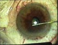
Currently, many devices and methods are available to provide both an adequate pupil intraoperatively and a functionally and aesthetically acceptable pupil postoperatively (Table 2). The easiest method to achieve a desirable pupil is to stretch the pupil bimanually with either2 separate push-pull instruments or prong-like devices specifically designed to stretch the pupil sphincter.3 Stretching the pupil margin may cause multiple fine partial sphincter tears, but the tears rarely create a deformed pupil after surgery. Infrequently, however, pupil stretching may induce an aesthetic change. Slide 4 shows an eye with an iris hematoma that resulted from two-instrument stretching. Simple pupil stretch is accomplished under topical anesthesia with or without the addition of intracameral lidocaine. Additionally, the maneuver adds little to the total surgical time and virtually nothing to the cost of surgery. Pupil stretch should be performed under viscoelastic to maintain a deep anterior chamber to protect other anterior segment structures. It is best to push and pull the two instruments slowly, simultaneously, and fully across the chamber. When stretching is performed in this manner, tears of the sphincter are minimized. On the other hand, rapid, jerky stretching can result in more significant sphincter damage.

A stretch in one meridian with two instruments or with the Keuch (Katena, Denville, NJ) bipronged device may be adequate for most cases; however, occasionally, it is necessary to stretch in multiple meridians. This can be accomplished with a Beehler (Rhein Medical Inc., Tampa, Fla.) four-pronged device through the main cataract incision or by working through multiple paracenteses with separate push-pull instruments. Another method to enhance the effect of the pupil stretch is to make a series of mini sphincterotomies with either Vanass scissors through the cataract incision or with vitreoretinal scissors placed through the paracentesis. Additional stretching maneuvers may then be performed with an improved likelihood for success.4 If only very small incisions are made in the sphincter tissue, the pupil will function normally and will be aesthetically appealing after surgery as noted in Slide 5.
Commercially available iris hooks (Grieshaber, Schaffhausen, Switzerland) have been designed by De Juan for use in complex posterior segment surgery (Slide 6).5 These hooks have been used during cataract surgery but must be used with caution to prevent iris damage during phacoemulsification.6,7 Slide 7 demonstrates that the hooks can anteroplace the iris diaphragm with resultant chaffing and thermal damage during phacoemulsification. Slide 8 reveals iris damage in the eye of a patient with a miotic pupil and an extraordinarily dense cataract that required a prolonged emulsification while iris hooks were in place. Iris damage may be avoided if the hooks are positioned parallel to the iris plane through small, short tract, peripheral paracenteses or by releasing the hooks after creating the capsulorrhexis but before phacoemulsification. Furthermore, the hooks should be used to create an adequate size pupil rather than a very large pupil to prevent the occurrence of an atonic pupil postoperatively. Similar to pupil stretch methods, gradual enlargement of the pupil is preferred over rapid, sudden tugging. While iris hooks may add to the expense and time of surgery, they are valuable for a surgeon who requires a relatively large pupil and may be used if a pupil should become miotic intraoperatively. An innovative use for the hooks has been to stabilize a loose capsular bag during surgery by placing the hooklets around the edge of the anterior capsulorrhexis.
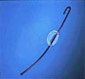
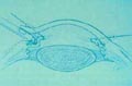
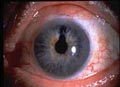
Another class of devices has been designed to enlarge the miotic pupil without damaging the sphincter (Table 2). Incomplete pupil ring expanders are placed in the anterior chamber through the main incision and manipulated into the pupil space. These expanders represent the most recent addition to the tools for surgical management of a cataract in the presence of a miotic pupil and are made of solid PMMA, silicone, or expansible hydrogel material. Although the expanders add to the cost of the surgery, they can create the largest diameter pupil with the least tendency for sphincter tears, without placing point pressure on the pupil margin. Rather, they enwrap the sphincter for approximately 300º. Although these devices may be cumbersome and time consuming to place properly, and may interfere with the progress of phacoemulsification, they are likely to be improved and may become commonly used, because of the generous pupil that they afford. Additionally, care must be taken to disengage the device from the pupil when removing it from the eye. Irrespective of the chosen method for enlarging the pupil during phacoemulsification, the pupil should be constricted at the close of surgery to prevent capture by the optic, adhesion to the capsule, or other manner of deformity.
References
- Guzek JP, Holm M, Cotter JB, et al. Risk factors for intraoperative complications in 1000 extracapsular cataract cases. Ophthalmology. 1987;94:461-466.
- Masket S. Relationship between postoperative pupil size and disability glare. J Cataract Refract Surg. 1992;(18):506-507.
- Shephard DM. The pupil stretch technique for miotic pupils in cataract surgery. Ophthalmic Surg. 1994;24:851-852.
- Fine IH. Pupilloplasty for small pupil phacoemulsification. J Cataract Refract Surg. 1994;20:192-196.
- De Juan Jr E, Hickingbotham D. Flexible iris retractors. Am J Ophthalmol. 1991;111:766-777.
- Nichamin LD. Enlarging the pupil for cataract extraction using flexible nylon iris retractors. J Cataract Refract Surg. 1993:19:793-796.
- Masket S. Avoiding complications associated with iris retractor use in small pupil cataract extraction. J Cataract Refract Surg. 1996;22:168-171.