Introduction
Beginning with the silicone taco lens, many ophthalmologists predicted that foldable IOLs were simply a flash in the pan and not going to have any lasting impact. Early on, people reported that some of these lenses were turning brown and predicted that the lenses would all become cataractous with time.1 Others reported marked difficulty in performing a YAG laser posterior capsulotomy2 and a significant increase in capsular opacification and inflammation in the eye. Add to this the general concept that the decrease in wound size over PMMA was a short-term difference at best, and it is understandable why so many people felt that PMMA would always be the best choice. The longevity of foldable lenses at birth seemed tenuous at best.
The browning of IOLs and the thought that they may become opaque with time dramatically impacted the sales of silicone IOLs until this problem was resolved (Slide 1). It is important to point out that, although a good series has never been published, the lenses that showed a brown haze were never more than a few percent of production lenses at any time. Although progression was noted, this generally peaked 3 to 6 months after surgery and did not worsen after that time. Manufacturers realized that the browning was simply water making its way into the silicone optic, which, once a steady state was reached, did not progress. Studies were performed to determine the impact of browning on visual acuity, contrast, and glare disability. There was no clinical effect. Unfortunately, none of this was published. Still, it was believed that the problem was one best to control and, therefore, through quality control processes and, in particular, complete material polymerization, the issue has been relegated to a dusty chapter in the past.
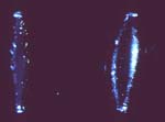
The YAG capsulotomy problem showed silicone to be much more sensitive to pitting and marking than PMMA. 2 Concerns raised about that issue were significant. However, appropriate understanding of new techniques for capsulotomy with this lens has resolved this issue as well. Approaches such as using less power with subtle defocus, as well as improved Nd:YAG laser machines, have helped. Other techniques, such as avoiding the optical center completely and making the capsulotomy only in the periphery, have solved this problem. Although sensitivity to YAG damage is increased for silicone lenses, those experienced in their use have not found this to be of clinical importance. No lens has been reported to be associated with significant laser pitting, decreased visual acuity, or other visual problems.
Silicone oil is also a problem for silicone IOLs in that the view of the fundus can be severely degraded. This is less so for PMMA and hydrophobic acrylic IOLs and least for hydrophilic acrylic. Although this is a relative contraindication for silicone IOLs in the face of significant vitreoretinopathy, a solvent exists that eliminates the problem and should soon render this problem moot3 (Slide 2).
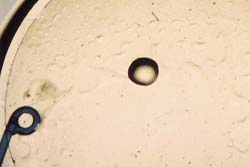

Looking at visual acuity, we were not surprised to find statistically better visual acuity without correction, knowing the astigmatic differences between the two eyes. However, we were surprised to see a statistically significant improvement in visual acuity with correction (Table 1). It was not apparent why there should be a difference between the two lenses in terms of best-corrected visual acuity, but we believed it must be related to posterior capsular opacification (PCO).
In a separate study looking at the same patients with 3-year follow-up, we looked subjectively and objectively at PCO.5 Our objective measurement was obtained by using a lens opacity meter and our subjective measurement by using standard photographs. We found that at 3 years the SI30 was statistically better than PMMA with regard to PCO. This explains the better best-corrected visual acuity (Table 2). PCO is a visual acuity problem as much as it is related to the complications associated with capsulotomy. Therefore, there are multiple reasons why capsulotomy should be avoided.
PCO
The history of PCO with foldable lenses is interesting. Plate lenses have shown promise in decreasing PCO without any randomized, prospective studies completed to date. It turns out that prevention of capsulotomy is most likely a possibility.6 The main concern has been posterior dislocation, which can occur after YAG capsulotomy due to the poor fixation of the haptics in the periphery of the bag. Although posterior dislocation is not a common problem, it is obviously disconcerting to both the surgeon and the patient. Therefore, plate haptic lenses used today require enlarged haptic holes to obtain better fixation. 7 Although animal studies clearly showed excellent fixation and larger holes have likely helped the incidence of posterior dislocation, this is still being reported. There are no prospective studies showing that larger holes have diminished the incidence of dislocation.
Three-piece silicone lenses have not been found to prevent PCO. The foldable lens, which from the beginning has been touted for its after cataract prevention, is a three-piece AcrySof IOL (Alcon Laboratories, Fort Worth, Texas). Emma J. Hollick, MD, and colleagues,8 examined the AcrySof, three-piece silicone, and PMMA and showed a statistically significant advantage of AcrySof in PCO prevention over both silicone and PMMA, with silicone and PMMA statistically the same. This, of course, contradicted our own study5 and remained a mystery until Hideyuki Hayashi, MD, released his prospective, randomized clinical trial9 looking at the SI30 three-piece silicone lens, AcrySof, and PMMA. With 2 years of follow-up, he showed that AcrySof is statistically much better than PMMA. However, interestingly, the SI30 and AcrySof were statistically indistinguishable in terms of YAG capsulotomy rates and objective measurement of PCO using Scheimpflug photography. Also, both were indistinguishable with regard to the improvement in visual acuity over PMMA, again showing that after cataract is more than just a YAG concern.
Further confirmation of the PCO prevention of the SI30 three-piece silicone IOL was presented in another study by Dr. Hollick and colleagues10 that looked at the SI30, PMMA, and a hydrophilic acrylic. Results showed that the PCO of the hydrophilic acrylic was significantly worse than PMMA and the SI30 significantly better than PMMA (Table 3). Unfortunately, they did not compare this lens in this prospective study with AcrySof. Studies presented by Kohnen11 and Kapainen12 have also shown equivalency between AcrySof and the SI40 IOL, which is basically an SI30 IOL with PMMA rather than polypropylene haptics. All of this suggests that the second-generation silicone lens has some distinct advantage over first-generation silicone with results comparable to AcrySof. To date, the only studies that have shown AcrySof superior to the SI40 in PCO prevention are a study by Ernest,13 and the autopsy series by Apple.14 The results of Ernest's study are still impressive for both lenses. However, the researchers have a follow-up of more than 4 years. AcrySof is clearly doing better with regard to PCO prevention.
Knowing that an IOL has a superior PCO profile is only part of the equation. We must also answer the question of why. Work by Okihiro Nishi, MD, has been instrumental in understanding some of the factors that have resulted in PCO prevention. He has been most interested in the barrier effect created by the lens, which creates a fold in the capsule (a discontinuous bend) mechanically blocking lens epithelial cell (LEC) ingrowth and proliferation under the IOL. His thesis has been that the truncated edge of AcrySof is the reason it has shown excellent PCO prevention. In support of this theory, he has shown that a truncated edge on PMMA in an animal model was equal in PCO prevention to AcrySof15 and that AcrySof with a rounded off edge did not prevent PCO. A silicone IOL with a truncated edge was also successful in blocking PCO.16 He further theorized that early postoperative posterior pressure on the part of the lens created by anterior capsular contraction is an important part of this process. In addition, he showed that the SI30/SI40 profile had the greatest posterior movement.17 Possibly, the SI30/SI40 lenticular design with a peripheral flange is an important part of this PCO prevention. Also good, but intermediate in early posterior movement, was hydrophobic acrylic and PMMA with no posterior movement for hydrophilic acrylic. Clearly, these biomechanical factors are the best elucidated and most important factors to be used in IOL manufacturing for PCO prevention.
Rupert Menapace, MD,18 presented his work showing that any breach in the barrier effect of the edge will eventually result in PCO. This would include wrinkles in the posterior capsule or a capsulorrhexis that does not completely cover the edge of the optic. An IOL with an incomplete 360° posterior edge such as single-piece AcrySof is another example of how PCO might start.
A biofilm theory has also been proposed to explain why the AcrySof lens has had such a low PCO rate. This theory basically relates to a sandwich of fibronectin between the capsule and a single layer of LECs with another layer of fibronectin between these LECs and the IOL, all of which produces a barrier to further lens epithelial cell growth.19 Clearly, AcrySof, in comparison to silicone and PMMA, has been shown to be more likely to produce such a biofilm.20 The downside to this theory is that it does not explain the biomechanical work of Nishi that looks at IOL edge profile in which the material itself has not had any impact on PCO. Also, it does not explain the clinical results from Hayashi's work9 and others, which does not suggest the importance of the material effect. Certainly, there is more to be known about the biofilm theory, and with time we will understand how important this might be in PCO prevention. The best way to find out about biofilm, however, will be to observe the one-piece AcrySof. The one-piece AcrySof does not have a complete 360° sharp edge profile due to the large haptics that extend to the posterior edge and the flaccid nature of the haptics, which regularly leave many wrinkles and folds without posterior IOL pressure in the early postoperative period. If biofilm alone is important, we would expect this lens to do well from a PCO standpoint; however, if a 360° discontinuous bend is more important, then this lens should not have the same PCO profile as the three-piece AcrySof IOL. I am not aware of any study to date that shows equivalency or a worsening of PCO with one-piece AcrySof; however, I am sure in the near future we will have the answer to this interesting question.
Hydrophilicity
Hydrophilicity is an interesting concept. The body certainly appreciates a substance that is hydrophilic, and, therefore, multiple studies show that hydrophilicity is an advantage. Other materials, such as hydrophobic acrylic and some silicone materials, have also done extremely well. However, hydrophilic materials always end up equal to or better than any other material with regard to inflammation. It is also true, however, that one way in which hydrophilicity shows its biocompatibility is the propensity for LEC growth on the anterior surface of the lens. This growth, which can occur minimally in hydrophobic acrylic, can be so exuberant as to completely cover the anterior surface of the lens, with some visual impact. The question is whether LEC growth anteriorly also results in LEC growth posteriorly with increased PCO.
In a PCO session at the 2000 European Society of Cataract and Refractive Surgeons meeting, four presentations involved a hydrophilic product, and in each case this product was statistically significantly worse compared to hydrophobic products with regard to PCO. None of this has been published; however, Yasar Küçüksümer, MD,21 compared the MemoryLens (CIBA Vision, Duluth, Ga.) to AcrySof with bilateral simultaneous implantation with 3 years of follow-up and showed a clinical and statistical difference in favor of AcrySof with regard to visual acuity, YAG capsulotomy, and PCO. At this point in time, hydrophilicity appears to be a negative factor with regard to PCO; however, other biomechanical factors engineered into the lens could possibly obviate some of the negative biomaterial factors associated with hydrophilicity.
The issue of hydrophilicity now involves other concerns that have recently been reported. Probably the most frightening effect to date has been the calcification of Hydroview IOLs (Bausch & Lomb, Claremont, Calif.) with reports from around the world. Although there are several hundred cases of calcification reported at this time and it represents only a small percentage of all lenses, it is a complication that we have not previously seen. Calcification tends to progress slowly and can be significant in some patients even to the extent that the lens must be removed.22 Although there is no definitive explanation, the manufacturers have suggested that a breakdown in the blood-iris barrier caused by silicone particles from the packaging may be the culprit. Nothing is officially published on this subject, and I consider the theory not proven to the extent that I have been able to review it. We must keep a careful eye on Hydroview lenses with regard to this calcification issue because the problem occurs early, and it may increase with time and become a significant problem. A more pertinent question is whether calcification is a potential problem with all hydrophilic materials. Fortunately, the only lens that has shown this tendency to date is the Hydroview lens.
The MemoryLens was also associated with cases of unusual inflammation, which can lead to toxicity. Our group published a series of cases demonstrating this unexplained inflammation, which fits into the rubric of toxic anterior segment syndrome (TASS).23 The manufacturer suggested that the inflammation was due to a residual polishing compound on the lens. Consequently, the MemoryLens was recalled from the market. The fact that inflammation can result in toxicity has been known for a long time, but it is something we have not seen for many years. I suggest that hydrophilicity, although a positive feature from some standpoints, may be a manufacturing nightmare due to the need to keep the lens dehydrated to the extent that the usual solvents that are used to clean hydrophobic lenses cannot be used. Therefore, from a manufacturing standpoint, there is a new, different, and probably more difficult level of quality control necessary to ensure the absence of any substance that may cause inflammation. Guaranteeing optical consistency presents another challenge. Our group has completed a study24 showing a significant decrease in refractive accuracy with the MemoryLens. The lens will be re-released with the assurance that the issues have been resolved; however, we must look carefully at hydrophilic materials for outbreaks of inflammation and refractive stability as other potential concerns. I am confident that manufacturers will, however, eliminate these problems with time.
Collamer is a new hydrophilic material just recently released that has shown some interesting properties. The U.S. Food and Drug Administration (FDA) approval application showed excellent results. The lens, when hit with an Nd:YAG laser, will self-heal the defect.
To date, there have been no reports of unusual complications and it is too soon to know what its PCO profile may be. Its hydrophilicity and shape suggest that PCO will be no better and most likely worse than PMMA; however, only time will tell. We should not forget the law of unintended consequences with all materials, and it is only prudent to have a heightened level of concern with any new class of materials until we have years of experience.
Investigation
Although all approved IOLs have good inflammation profiles, clinical differences have been shown to exist. Hollick, in looking at giant cell formation on lenses, showed AcrySof to be quieter than PMMA and silicone, which were similar to each other.25 Guiseppe Ravalico, MD,26 however, showed that the SI30, a second-generation silicone IOL, is equivalent to hydrophilic acrylic with regard to the low amount of reactivity. This mystery was further clarified when Dr. Hollick compared the SI30 to hydrophilic acrylic and PMMA and showed that the SI30 and hydrophilic acrylic were both quiet and much better than PMMA.27 All of this certainly lends support to the differences between first-generation silicone and the SI30 and SI40 second-generation silicone IOLs. However, no direct comparison between this lens and AcrySof was reported until the work by Thomas Samuelson, MD (Table 4).
Samuelson performed a prospective study looking at SI30 and SI40, as well as AcrySof, first-generation silicone, and a plate lens, in glaucoma patients who had cataract surgery with IOL insertion and trabeculectomy.28 These triple procedures are particularly important in that with chronic small pupils, there often is increased inflammation and giant cell deposits that can be a clinically significant problem causing decreased vision and that must be removed with the YAG laser. At 6 months, Samuelson found approximately 5% of such deposits on the SI30 and SI40 IOLs with 15% on AcrySof lenses and 33% on the plate silicone lens. The plate silicone lens was statistically worse than AcrySof and the second-generation silicone SI30 and SI40. No similar studies have been performed to date to determine whether other second-generation silicones are comparable. Clearly, we have information that although the hydrophilic material uniformly will do extremely well, certainly, AcrySof and at least SI30 and SI40 second-generation silicone IOLs are either equivalent or close in their inflammatory profile. First-generation silicone seems to respond similarly15 to PMMA and significantly worse than the hydrophobic and hydrophilic acrylic and some second-generation silicone. The new Sensar IOL (Allergan) has not undergone sufficient clinical studies but it would appear to be similar to AcrySof in its inflammatory profile.
Unfortunately, many attributes of IOLs have proven to be double-edged swords. Although the flattened edge of AcrySof has clearly been important in preventing PCO, unwanted images have been shown to be a negative side effect. This first became apparent to me when I had a patient who had seen several ophthalmologists regarding debilitating unwanted images, particularly at night, that rendered his job at risk. I found that the patient had a perfectly centered MA30 AcrySof lens with the capsule for 360° overlapping of the IOL edge and 20/15 uncorrected vision. After discussion with the patient and having heard a few reports that this may be related to the lens, I was convinced that the patient was unable to function. We removed the lens and replaced it with an SI40 IOL, resulting in prompt relief of his symptoms. We have had several other such cases and combined our series with The University of California Los Angeles. Eight patients (nine eyes) required removal of the AcrySof lens for debilitating symptoms. Their symptoms were either completely or largely resolved with a round-edge IOL.29
With unwanted images now well documented, Holladay and colleagues30 showed through ray-tracing studies that the truncated edge creates a mirror effect and can result in a central flash even if the patient is not looking directly at an offending light. Patients also complain of the reflective pupil associated with the high refractive index of AcrySof and its relatively flat anterior curvature. Some patients complain about a shimmering quality to their vision. James A. Davison, MD,31 also reported negative photopsia of grainy gray temporal vision, which can be severe enough to necessitate removal of the lens. We reported an outcomes study32 in which unwanted images were a common complaint. A central flash at night was a common complaint for the AcrySof MA30 and MA60 lenses.
Fortunately, the industry has responded. The new one-piece AcrySof IOL has a frosted edge and the edge of the new three-piece AcrySof lens (MA60AC) has been changed to minimize these unwanted images. Both of the newer AcrySof products also have an increased anterior curvature, which will minimize the glinting pupil. Any means to create a truncated posterior edge with the anterior rounded edge should also dramatically decrease this problem while maintaining the advantage of PCO prevention by the creation of a discontinuous bend in the capsule. An example is the new Sensar IOL with a modified edge, called the "Opti-edge."
There is still much that we do not know about unwanted images, and we must study them further to decrease this significant cause of symptoms in our otherwise satisfied patients. The industry, however, has advanced significantly from the state of the art of several years ago.
Our first experience with the AcrySof IOL was one of excitement as many of these lenses were implanted at our institution. We were surprised when, weeks after surgery, we found multiple, white glistening dots often loaded in the optic of the IOL. These have been called glistenings and were a surprise in severity, not only to us, but also to the manufacturer. The manufacturer realized that this occurred due to a change in packaging. The AcryPak packaging was the cause of these glistenings because the older lenses in the Wagon Wheel configuration did not have this problem.
We reported that severe glistenings in patients who had a silicone lens in one eye and AcrySof in the other resulted in a statistically significant loss of contrast sensitivity, but no significant loss of visual acuity, although our power with only 10 patients studied was low.33 We performed an in vitro study that showed at 37° C, glistenings occurred only with the AcryPak packaging and not with the Wagon Wheel packaging.34 Serendipitously, we discovered that AcrySof, in either the Wagon Wheel or AcryPak packaging, after cooling to room temperature, showed a marked increase in the incidence of glistenings. These glistenings could reliably be produced and just as reliably disappear by warming the lens to 37° C. Still, these acute glistenings in our patients improved, resolving slowly with time, so we believed that this issue was of no significant concern.
Not until the 1999 American Society of Cataract and Refractive Surgeons meeting, when Katsuya Mitooka, MD,35 presented a poster, did we have renewed concerns about AcrySof. Mitooka showed that glistenings in AcrySof were starting to appear over time, often years after implantation, with a significant loss of contrast sensitivity and increased severity associated with diabetes. H. Burkhard Dick, MD, suggested that a breakdown of the blood-iris barrier and increased protein in the eye that is seen with diabetes results in increased severity of glistenings. This theory was confirmed by an in vitro study in which serum was added to the fluid surrounding incubated AcrySof IOLs.36 This combination of work prompted us to review our own patients in a pilot project37 including at least 10 patients each year from 1 to 4 years with 42 patients studied to look at glistenings. Although we did not prove progression with time, the results were highly suggestive (P = 0.06). We found approximately 10% with significant glistenings and with careful examination, all lenses had some glistenings. Although we found no statistically significant difference in contrast sensitivity looking at the more severe glistenings compared with the remainder of the patients, we found a small but statistically significant loss of visual acuity. This suggests that there is some clinical significance to glistenings and not just a cosmetic concern (Table 5).
The advent of new acrylic material prompted an in vitro temperature stress test study of three-piece AcrySof, one-piece AcrySof, and Sensar, which was a new hydrophobic acrylic that had not been shown to have glistenings in its clinical history.38 We placed these lenses in cells of balanced salt solution and monitored them at 37° C each day for 5 consecutive days decreasing the temperature to room temperature for 2 hours each day and then viewing the changes in glistening formation. We found a low level of glistenings in all three materials at 37° C with stability and equivalency among the three IOLs. Lowering the temperature of the lenses to room temperature, however, resulted in a dramatic increase of glistenings in the one-piece AcrySof lens. This amount of glistenings was too high to count. Interestingly, three-piece AcrySof IOLs responded differently with an actual decrease in countable glistenings by a slit lamp examination at room temperature. This was associated with an increase in haze. At a magnification of 80 times, innumerable glistenings averaging 4 µm in diameter were detected.
The Sensar was the most stable of the IOLs examined, but we did see a faint bluish haze at room temperature with lucid areas. At the center of each lucid area would be a single relatively large glistening. Fortunately, all lenses responded with stability and elimination of this acute change when returned to 37° C, with each day relatively consistent throughout the 5 days for all three lenses. We concluded that the biophysical nature of these lenses is different, even though one- and three-piece AcrySof IOLs are the same material. Whether there will be a clinical difference between one- and three-piece AcrySof remains to be seen and whether glistenings will be an issue at all in the Sensar is also unknown. However, this study showed differences, and it will be interesting to see how the study results correlate with the clinical situation over time.
Dr. Wilkins contacted me with regard to small white spots that he was seeing in PMMA IOLs, which prompted a thorough evaluation of all of his patients who had a Surgidev B20-20 IOL (Advanced Vision Science, Goleta, Calif.). This lens is interesting because it was mold injected and, therefore, has a very low molecular weight, which has been shown in the past to be associated with some glistenings and also sensitivity to YAG laser damage.39 With an average of more than 10 years of follow-up, Wilkins found that an overall incidence of 89% of these IOLs showed glistenings that appear to be analogous to the situation in AcrySof. We clearly documented that glistenings are worsening with time. All lenses followed for more than 5 years showed glistenings and there is no obvious end point that can be determined at this time. Some lenses took more than 10 years before any glistenings appeared.40
My colleagues and I have seen that water in any of the hydrophobic IOL materials can result in either the browning associated with silicone IOLs or glistenings in hydrophobic acrylics and PMMA. We have had enough experience with PMMA to suggest that other than this mold injected lens, which is not how lenses are produced today, and in AcrySof lenses, there is no evidence of these side effects being clinically important. It would seem best to eliminate this problem because some clinical impact has been documented. With evidence of progression over time, we do not know exactly what the end stage is for these lesions. The PMMA study shows that we cannot assume any material is free of glistenings without at least 10 years of follow-up.
We began this study of foldable IOLs because we were interested to see what the smaller wound might do with regard to astigmatism and its stability, as well as visual acuity, over time. Now that we have many foldable lenses, it is apparent that they are not equivalent with regard to wound size. Therefore, this raises a question of whether wound size variability has an impact on astigmatism or wound stability. James Memmen, MD, using go, no-go gauges, designed by Jim Deacon, MD, evaluated a host of foldable lenses.41 Memmen found that the smallest measured incision size for insertion of the SI55 using the Unfolder (Allergan) averaged 2.65 mm. The largest incision used for the insertion of the MA60 AcrySof was 3.9 mm. The incision used for the SI30 and SI40 was approximately 2.9 mm. Knowing whether this variation in wound size has impact on astigmatism would require a long prospective study.
Looking at wound stability, we could see it would be a fairly easy project, and, therefore, we decided to look at 200 consecutive surgeries performed with the MA60, which is a common lens at our institution, and 200 consecutive cases done with the SI40 and the Unfolder. The significant difference is that the SI40 with the Unfolder easily goes through the phacoemulsification wound without enlargement, whereas the MA60 must have the wound enlarged with a final wound size of approximately 4.0 mm. We evaluated each patient's chart for evidence of wound instability, which we defined as a need to suture the wound in the operating room or on the first postoperative day due to a wound leak.
Our evaluation found a statistical difference with 13 wound events out of 200 eyes (6.5%), with the MA60 and only three wound events (1.5%) with the SI40 (P = 0.01).42 There was greater stability with the smaller incision and less wound events, although none of them resulted in serious long-term complications. It is preferred to be able to put the foldable lenses through an unenlarged phaco incision.
Conclusion
All of these studies to date have been exciting in showing that foldable IOLs have distinct advantages with regard to their wound size allowing greater wound stability and decreased astigmatism that can be shown to make a difference more than 3 years after surgery. Additionally, we now have lenses that have distinct advantages with regard to decreased PCO and an improved inflammatory profile, all of which can be obtained without an increase in unwanted images or glistenings in the lens. More exciting is that we now understand the principles, such as the posterior truncated edge, which will help manufacturers continue to improve the product. The market has responded to foldable lenses and these lenses are clearly the dominant force in the United States and Europe. Although this evolution has occurred, the room for improvement is evident and we expect even better products in the future.
References
- Milauskas AT. Silicone intraocular lens implant discoloration in humans [letter]. Arch Ophthalmol. 1991;109:913-915.
- Bath PE, Boerner CF, Dang Y. Pathology and physics of YAG-laser intraocular lens damage. J Cataract Refract Surg. 1987;13:47-49.
- Hoerauf H, Menz DH, Dresp J, Laqua H. Use of 044 as a solvent for silicone oil adhesions on intraocular lenses.
J Cataract Refract Surg. 1999;25:1392-1396.
- Olson RJ, Crandall AS. Prospective randomized comparison of phacoemulsification cataract surgery with a 3.2 vs a 5.5 mm sutureless incision.
Am J Ophthalmol. 1998;125:612-620.
- Olson RJ, Crandall AS. Silicone versus polymethylmethacrylate intraocular lenses with regard to capsular opacification.
Ophthalmic Surg Lasers. 1998;29(1):55-58.
- Olson RJ. Consultation Section: Cataract surgical problem.
J Cataract Refract Surg. 1999;25:1426-1427.
- Mamalis N, Omar OH, Viega J, Tanner DS, Fernquist D. Comparison of two plate haptic intraocular lenses in a rabbit model.
J Cataract Refract Surg. 1996;22(52):1291-1295.
- Hollick EJ, Spalton DJ, Ursell PG, et al: The effect of polymethylmethacrylate, silicone and polyacrylic intraocular
lenses on posterior capsule opacification three years after cataract surgery. Ophthalmology.
1999;106:49-55.
- Hayashi H, Hayashi K, Nakao F. Quantitative comparison of posterior capsule opacification after polymethylmethacrylate,
silicone and soft acrylic intraocular lens implantation. Arch Ophthalmol. 1998;116:1579-1582.
- Hollick EJ, Spalton DJ, Ursell PG, Meacock WR, Barman SA, Boyce JF. Posterior capsular opacification with hydrogel,
polymethylmethacrylate, and silicone intraocular lenses: two-year results of a randomized prospective trial.
Am J Ophthalmol. 2000;129:577-584.
- Kohnen T. Posterior capsule after silicone and hydrophobic acrylic IOL implantation. Presentation at the
American Society of Cataract and Refractive Surgery, Boston, MA, May 20, 2000.
- Kaipiainen S. Comparison of YAG capsulotomy rates between PhacoFlex II and AcrySof IOLs after simultaneous bilateral
implantation. Presentation at the American Society of Cataract and Refractive Surgery, Boston,
MA, May 24, 2000.
- Ernest P. PCO in eyes with AMO PhacoFlex II and Alcon AcrySof MA30BA IOLs. Presentation at the
American Society of Cataract and Refractive Surgery, Boston, MA, May 20, 2000.
- Apple DJ, Peng Q, Visessook N, Werner L, Pandey SK, Escobar-Gomez M, Ram J, Auffarth GU. Eradication
of posterior capsule opacification: documentation of a marked decrease in Nd:YAG laser posterior capsulotomy
rates noted in an analysis of 5416 pseudophakic human eyes obtained postmortem. Ophthalmology.
2001;108(3):505-518.
- Nishi O, Nishi K. Preventing posterior capsule opacification by creating a discontinuous
sharp bend in the capsule. J Cataract Refract Surg. 1999;25(4):521-526.
- Nishi O. The discontinuous capsular bend -sharp implant edge - rationale and experimental
evidence: 1999 European Society of Cataract and Refractive Surgeons, Vienna, Austria, 1999.
- Nishi O. Material vs. design: what really matters? Presentation at the American Society of
Cataract and Refractive Surgery, Boston, MA, May 23, 2000.
- Menapace R. Material vs. design: what really matters? Presentation at the American Society of
Cataract and Refractive Surgery, Boston, MA, May 23, 2000.
- Linnola R. Sandwich theory: bioactivity-based explanation for posterior capsule opacification.
J Cataract Refract Surg. 1997;23(10):1539-1542.
- Linnola RJ, Werner L, Pandey SK, Escobar-Gomez M, Znoiko SL, Apple DJ. Adhesion of fibronectin,
vitronectin, laminin, and collagen type IV to intraocular lens materials in pseudophakic human autopsy eyes.
Part 1: histological sections.
- Küçüksümer Y, Bayraktar S, Sahin S, Yilmaz ÖF. Posterior capsule opacification 3 years after
implantation of an AcrySof and a MemoryLens in fellow eyes. J Cataract Refract Surg.
2000;26:1176-1182.
- Werner L, Apple DJ, Escobar-Gomez M, Ohrstrom A, Crayford BB, Bianchi R, Pandey SK.
Postoperative deposition of calcium on the surfaces of a hydrogel intraocular lens.
Ophthalmology. 2000;107(12):2179-2185.
- Jehan FS, Mamalis N, Spencer TS, Fry LL, Kerstine R, Olson RJ. Postoperative
sterile endophthalmitis (TASS) associated with MemoryLens. J Cataract Refract Surg.
2000:26:1773-1777.
- Nelson T, Zabriskie NA, Brodstein DE, Baker MR, Alder SC, Richards BW. Significant
postoperative refractive errors in vivo with the Mentor MemoryLens intraocular lens implant.
Submitted to J Cataract Refract Surg.
- Hollick EJ, Spalton DJ, Ursell PG, Pande MV. Biocompatibility of poly (methyl methacrylate),
silicone, and AcrySof intraocular lenses: randomized comparison of the cellular reaction on the
anterior lens surface. J Cataract Refract Surg. 1998;24:352-360.
- Ravalico G, Baccara F, Lovisato A, Tognetto D. Postoperative cellular
reaction on various intraocular lens materials. Ophthalmology. 1997;104:1084-1091.
- Hollick EJ, Spalton DJ, Ursell PG. Surface cytologic features on intraocular lenses: can increased
biocompatibility have disadvantages? Arch Ophthalmol. 1999;117:872-878.
- Samuelson TW, Chu YR, Kreiger RA. Evaluation of giant-cell deposits on foldable intraocular
lenses after combined cataract and glaucoma surgery. J Cataract Refract Surgery. 2000;26(6):817-823.
- Farbowitz MA, Zabriskie NA, Crandall AS, Olson RJ, Miller KM. Visual complaints of patients with AcrySof acrylic
intraocular lenses. J Cataract Refract Surg. 2000;26:1339-1345.
- Holladay JT, Lang A, Portney V. Analysis of edge glare phenomena in intraocular lens edge designs.
J Cataract Refract Surg. 1999;25(6):748-752.
- Davison JA. Positive and negative dysphotopsia in patients with acrylic intraocular lenses.
J Cataract Refract Surg. 2000;26:1346-1355.
- Tester R, Olson RJ, Pace NL, Samore M. Dysphotopsia in phakic and pseudophakic
patients--incidence and relation to intraocular lens types. J Cataract Refract Surg. 2000;26:810-816.
- Dhaliwal DK, Olson RJO, Mamalis N, et al. Visual significance of
"glistenings" seen in the AcrySof lens. J Cataract Refract Surg. 1996;22:452-457.
- Omar OH, Pirayesh A, Mamalis N, Olson RJ. In vitro analysis of AcrySof intraocular lens
"glistenings" in AcryPak and wagonwheel packaging. J Cataract Refract Surg. 1998;24:107-113.
- Mitooka K, Hiroshi T, Takuya S. Glistening in acrylic IOLs. ASCRS Symposium on Cataract,
IOL, and Refractive Surgery, Seattle, Washington, April 10-14, 1999.
- Dick HB, Olson RJ, Schwenn O, Magdowski, Pfeiffer N. Vacuoles in the AcrySof intraocular
lens as factor of the presence of serum in aqueous humor. Ophthalmic Research. 2001;33(2):61-67.
- Christiansen G, Durcan FJ, Olson RJ, Christiansen K. Prevalence, severity, and visual significance of
glistenings seen in the AcrySof IOLs one to four years from implantation. Accepted by J Cataract Refract Surg.
- Gregori N, Spencer T, Mamalis N, Olson RJ. In Vitro comparison of hydrophobic acrylic intraocular lenses in regard
to glistening formation. Submitted to J Cataract Refract Surg.
- Wilson SE, Brubaker RF. Neodymium: YAG laser damage threshold. Ophthalmology. 1987;94:7-11.
- Wilkins E, Olson RJ. Long-term glistenings with the Surgidev B20/20 PMMA intraocular lens. Accepted by
J Cataract Refract Surg. 2001.
- Memmen JE. The cataract wound, how big is it, really? Royal Hawaiian Eye Meeting; Hawaii,
January 24-29, 1999.
- Kumar R, Reeves DL, Olson RJ. Wound complications associated with incision enlargement when placing foldable
intraocular lenses during cataract surgery. J Cataract Refract Surg. 2001;27:224-226.