Introduction
Presbyopia
Accommodation is the process that allows the human crystalline
lens to change its focus from far to intermediate or near vision.
With age, the crystalline lens loses its ability to focus on near
objects, causing eye strain and fatigue, blurred vision, and
headaches. This condition, known as presbyopia, begins in
childhood. It affects the majority of people in their 40s and
affects everyone by age 50.
Theories on Accommodation
There are several theories on the mechanism of accommodation.
The most widely accepted theory is that of Hemholtz proposed
in 1855. Hemholtz believed that the hardening of the natural
crystalline lens causes the loss of accommodation. It is known
that unlike the skin, the lining of the digestive tract and bladder,
where the dead ectodermal cells can be shed to the exterior of the
body, the ectodermal cells that make up the natural crystalline
lens cease their mitotic activity and are retained in the capsular
bag. It is theorized that the continuous production of new cells
within the lens capsule causes the crystalline lens to harden,
enlarge, and lose its elasticity. These changes in the crystalline
lens prevent it from changing shape to provide near vision,
that is,
the ability to accommodate is lost.
Numerous scientific studies of accommodation have verified Hemholtz’s observations with few modifications. Studies by Adrian Glasser, PhD, a leading authority on accommodation, have reinforced the conclusion that lens hardening and enlargement must be considered as important factors in the development of presbyopia. Dr. Glasser further purports that with increasing age, the human lens gradually loses the ability to undergo optical changes in response to mechanical stretching.1-2 Despite the substantial experimental evidence to show that the human lens gets harder and larger with increasing age, the classical views of loss of accommodation with age have been refuted by Ronald Schachar, MD,3 who has developed a theory that presbyopia is not primarily due to hardening of the lens, but is caused by increased equatorial growth of the lens. However, Schachar’s theory is controversial and is currently under study in the United States.
Current Technologies for the Treatment of Presbyopia
There are currently two approaches to the management of
presbyopia: surgical and nonsurgical. The most commonly used
nonsurgical treatment consists of bifocal reading glasses, which
were invented by Benjamin Franklin in the 1780s. The bifocal
spectacle enables presbyopes to see at two fixed distances
(distance and near); however, modern day consumers seek more
cosmetically acceptable solutions. The advent of the soft contact
lens in 1961 dramatically expanded the number of patients
wearing contact lenses. These lenses have been used as a
platform for developing solutions for presbyopia, including multifocal contact lenses.
In contrast to bifocal spectacle lenses, multifocal contact lenses focus multiple images onto the retina. The brain then selects the optimally focused image, with the selection depending upon the object being viewed. Over time, some patients are able to achieve comfortable near and distance vision. However, many patients are unable or unwilling to complete the process of “learning” image selection which is required for the successful use of multifocal contact lenses. In addition, glare, particularly at night, is a significant complaint. Consequently, this technology has not met with widespread acceptance.
Monovision is a technique that can be used with either contact lenses, intraocular lenses or the commonly performed vision correction procedure, laser in situ keratomileusis (LASIK). In monovision, one eye is corrected for near vision (using a contact lens, intraocular lens, or LASIK), and the other eye is focused at distance. Some patients complain of blurred vision, glare with difficulty driving at night and loss of depth perception and are unable to tolerate these side effects.
Another alternative for the correction of presbyopia, still considered investigational, is a scleral expansion surgical procedure that operates on the principles developed by Ronald Schachar, MD. However, results have been mixed, and only limited data are available from prospective clinical trials.
Intraocular Lenses
Conventional intraocular lenses, which focus light at a fixed
distance, offer excellent visual outcomes to the majority of
patients undergoing cataract extraction using modern surgical
techniques. For near vision, most patients require reading glasses
or bifocals following cataract surgery since intraocular lenses
provide a single focal distance. Intraocular lenses that provide
multiple focal distances for both near and distance vision, i.e., multifocal lenses, offer an alternative to patients seeking
pseudophakic presbyopia correction. Because multifocal
intraocular lenses provide more than one focal distance
simultaneously, the optical quality of the retinal image is worse
than that produced with a monofocal lens. Many patients in
particular complain of halos at night. This loss of image quality
can affect visual performance and has led to interest in
alternative modalities for presbyopia correction in pseudophakes
following cataract surgery.
The role of the Ciliary Body in Accommodation
Filling of the capsular bag with inflatable endocapsular balloons
or in situ cured silicone polymeric gels has been evaluated in
animal models to determine whether the accommodative
properties of the natural crystalline lens can be replicated.4-7 Such
studies presuppose the continued functionality of the ciliary
muscle, such that removal of a cataractous crystalline lens
allows the ciliary muscle to respond to accommodative stimuli.
While there have been conflicting theories on whether the ciliary
muscle atrophies with age, it has been shown in a series of
published reports that the ciliary muscle not only maintains its
strength through the aging process but experiences an increase in
force.8-11 This was demonstrated in a study designed to evaluate
the ability of an artificial lens to restore accommodation in the
senile eyes of rhesus monkeys, a species which experiences the
presbyopic loss of accommodation.4,12-14 In this study, the
decrease in anterior chamber depth was significantly higher in
the operated eyes of each rhesus monkey than in the contralateral
control eye, which had not been implanted and failed to
demonstrate any accommodative change. Recent studies using
magnetic resonance imaging (MRI) have shown only a slight loss of ciliary muscle contraction with increasing age in patients 80 years of age
or older.15
J. Stuart Cumming, MD, through clinical observation, noted that a small number of his cataract patients were able to see at near through their distance correction after implantation with a plate-haptic intraocular lens. Plate-haptic lenses are located against the vitreous face, and have the ability to forward flex slightly. This observation suggested that there was an apparent ability of the ciliary muscle to move a plate-haptic intraocular lens forward and prompted Cumming to appreciate that even in presbyopes, the ciliary muscle may maintain some function and have the ability to move the optic of a carefully designed plate-haptic intraocular lens forward upon constriction. This would enable the patient to see at near.
Birth of the Accommodating Intraocular Lens
Accommodation and Pseudoaccommodation
It is known that a certain proportion of patients implanted with
standard looped monofocal intraocular lenses appear to have
functional near vision without glasses following lens
implantation. This effect can be attributed to either
accommodation, pseudoaccommodation, or a combination of
both. Accommodation would be the result of the optic of the
intraocular lens moving forward upon constriction of the ciliary
muscle. In 1958, Spencer Thornton demonstrated by A-scans that
the optic of a three-piece intraocular lens could move forward
during constriction of the ciliary muscle in some patients.16-17
Pseudoaccommodation would be due to one or a combination of
the following: some degree of myopia, an aspheric cornea
(i.e., steep at 90º), or a small pupil, resulting in increased
depth of focus.18-22
A literature search revealed that the ciliary muscle, upon constricting, undergoes a redistribution of its mass with encroachment into the vitreous cavity. This could provide vitreous pressure against the posterior lens surface, possibly inducing forward movement of an intraocular lens23 (Slide 1). In 1958, Jackson Coleman demonstrated a decrease in pressure in the anterior chamber with a simultaneous increase in pressure in the vitreous cavity upon stimulation of the ciliary muscle in 10 primates24 (Slide 2).
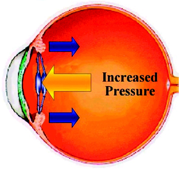
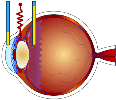
In a population of patients implanted with silicone plate lenses, when near vision was measured through the distance correction, at the phoropter, effectively eliminating the pseudoaccommodative effects of myopia, small pupil size, and minus cylinder, a number of patients continued to have good near vision.
This suggests that the action of the ciliary muscle alone may have been responsible for the ability of these patients to see at near (Cumming, unpublished data).
This hypothesis was further explored by performing A-scan measurements on these few patients who had near vision presumably resulting from action of the ciliary muscle. A-scan measurements were made following cycloplegia with tropicamide and compared to A-scans performed following the administration of pilocarpine. In a group of 10 patients with good distance-corrected near vision, A-scan measurements of the vitreous cavity were approximately 0.7 mm longer following pilocarpine administration than when performed after administration of tropicamide. These data serve to support the ability of the ciliary muscle to move a silicone plate-haptic lens forward in a few, carefully selected patients, as evidenced by the increased length of the vitreous cavity after treatment with pilocarpine. Further, since the other elements associated with pseudoaccommodation (i.e., myopia small pupil, minus cylinder) were eliminated as factors in providing near vision in these patients by measuring corrected near acuity at the phoropter, it appeared that near vision may have resulted from action of the ciliary muscle on the vitreous body and the lens (Cumming, unpublished data).
These findings are consistent with data reported by Hardman Lea and colleagues25 and by Niessen and colleagues.26 In two similar studies of pseudophakic accommodation, anterior chamber depth was decreased following pilocarpine administration, indicating forward movement of the intraocular lens. While there was a significant difference in anterior chamber depths with pilocarpine as compared to cyclopentolate, the magnitude of this difference was reported to be approximately 0.2 mm to 0.25 mm, considerably smaller than the 0.7 mm change described above. Since it has been reported that silicone plate-haptic intraocular lenses have a propensity toward posterior positioning, or vaulting, in the capsular bag placing the optic up against the vitreous face,27-29 it can be postulated that the potential magnitude of forward movement of a plate-haptic lens is greater than that of a standard looped intraocular lens.
This is further supported by the results of a study comparing the vitreous cavity lengths pre- and postoperatively in eyes implanted with silicone plate-haptic lenses to those implanted with silicone multipiece looped lenses.27 In this study, six (50%) of the twelve eyes implanted with a plate-haptic lens had shorter vitreous cavity lengths postoperatively than preoperatively and all located 0.75 mm or less closer to the position of the posterior capsule prior to surgery. In the eyes implanted with silicone loop lenses, there was shortening of the vitreous cavity in only two of the 10 eyes (20%). This consistent posterior displacement of the plate lens optic places it up against the vitreous face in the capsular bag following implantation and fibrosis, which results in posterior vaulting of the uniplanar plate lens.
These studies confirm the continued strength and functionality of the ciliary muscle in elderly cataract patients and support the hypothesis that this muscle could be a primary force resulting in accommodation in pseudophakes.
Recent studies published in 1999 by Susan Strenk and colleagues30 demonstrated by high-resolution MRIs the redistribution of the ciliary body mass to invade the vitreous cavity upon accommodation. This confirmed the findings in the drawings made by Busacca in 1955 on an aniridic patient.23 These MRIs demonstrated ciliary muscle action in all ages including an 83-year-old patient.
Plate-Haptic Experience
The concept of an “accommodating” intraocular lens, a lens that
would take advantage of the continued functionality of the ciliary muscle after cataract removal, with resulting forward
movement of the optic to provide good, consistent near vision,
was developed by Cumming. The concept was based on his observations of
more than 2,000 implanted plate-haptic lenses. Cumming
had observed that these lenses always located in the posterior
part of the capsular bag space, which is 5 mm from front to back
immediately after cataract surgery before fibrosis takes place.27
This was demonstrated by measuring the vitreous cavity length
prior to and following cataract surgery after implantation of
silicone plate haptic lenses. Thus, the plate-haptic design
provides the maximal posterior positioning of the lens in the
capsular bag space. This would allow the greatest potential
forward movement of the lens upon constriction of the ciliary
muscle.
Silicone plate lenses with a 6-mm optic were shown to vault posteriorly in the capsular bag space in all of the implanted eyes studied, decreasing the vitreous cavity length in 50% of the eyes.27 Kammann and colleagues reported posterior positioning in a substantially greater proportion of eyes (21 of 22, or 96%) implanted with silicone plate lenses with a 5 mm optic.31 Since the overall haptic/optic length of the lenses in both of these studies was the same (10.5 mm), these data suggested that a smaller optic resulting in longer plate haptics may increase the likelihood of posterior vaulting of the lens with possibly greater potential forward movement of the optic upon ciliary body constriction.
As described above, the posterior position of silicone plate-haptic lenses in the capsular bag has been established through pre- and postimplantation measurements of vitreous cavity lengths. For optimal performance of a lens intended to move forward to provide near vision, maximal posterior positioning is desirable. Since the action of the ciliary muscle induces forward movement of the optic of the plate lens in the capsular bag, it was postulated that by eliminating the anterior-biasing force, maximum posterior positioning would be achieved. This was done by paralyzing the ciliary muscle during the early postoperative period when fibrosis of the capsular bag occurs. On this basis, administration of atropine 1% at the time of surgery and on the first postoperative day has been proposed as a means of ensuring the maximum posterior positioning and fixation of the lens in the distance focused and unaccommodated position up against the vitreous face.
Atropine has a long history of use in cycloplegia and dilation of the pupil by paralyzing the parasympathetic muscles. When applied topically to the eye, its effect lasts from 10-14 days. In addition to maximizing the posterior location of the accommodating intraocular lens, it prevents dislocation during the period of fibrosis and allows excellent centration. During the early studies of the initial accommodating lens models, one drop of atropine per day was applied to the eye for three weeks. Since patient compliance with this treatment regimen was a concern, a small sub-study was conducted to evaluate the need for atropine. The results of this study revealed that atropine administration could be reduced to 1 drop at the time of surgery and 1 drop on the first day after surgery. This regimen addresses potential patient compliance issues, since the surgeon or other personnel are responsible for the administration of atropine.
By allowing the lens to remain in the maximal posterior position, preventing forward movement during the period of fibrosis around the lens haptics, anterior vaulting could be avoided. It was also postulated that ciliary muscle action during the early postoperative period may be a cause of decentration and that centration would be facilitated by paralyzing the ciliary muscle.
It occurred to Cumming during his experience with implanting standard silicone plate-haptic lenses, that because these lenses are only 1.3 mm thick, there is a large space in front of the lens since the lens is about 5 mm thick in elderly patients. Therefore, a hinge could be placed across the plate haptic near its junction with the optic and the optic would have space to move anteriorly. The hinge was incorporated to facilitate the forward movement of the optic by minimizing the resistance to the possible pressure exerted on the lens by the forward movement of the vitreous body associated with contraction of the ciliary muscle.
Early Experience with the Accommodating Intraocular Lens
For the first accommodating lens, Cumming designed a
simple plate lens with a 4.5-mm optic and hinges. Two lenses
were implanted in England in 1991. One patient died within 3
months of surgery, but the other patient was found to have
functional near vision at a distance of 25 cm to 30 cm and an
accommodating amplitude of 3 D to 4 D by the fogging
technique. A-scans following the administration of pilocarpine
demonstrated more than 2 mm of anterior movement of the
optic. This result supports the design hypothesis that since the
plate-haptic lens locates so far posteriorly, a lens design was
possible where movement of approximately 1 mm could provide
up to 2 D of accommodation. It was also confirmed that
postoperative capsular fibrosis with anterior capsular constriction
play important roles in the successful functioning of the lens.
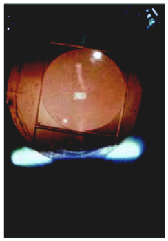
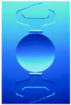
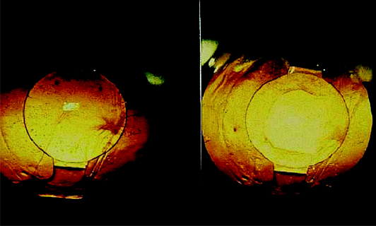
It was during this time that Cumming forged a collaboration with Professor Jochen Kammann, MD, in Dortmund, Germany. Kammann implanted a succession of six different accommodating intraocular lens designs, in a significant number of patients. The first two designs are shown in Slide 3A, Slide 3B, and Slide 4 Following implantation, accommodation amplitude was measured in two ways. First, patients were corrected to their best distance vision with a maximum plus refraction and examined in a dim room (to avoid the increased depth of focus created by a small pupil). Minus power was then added to the correction –0.25 D at a time. The endpoint of the accommodative amplitude was noted as the point immediately before patients lost one line of Snellen acuity. The second method was to perform a manifest refraction on the patient, administer pilocarpine to constrict the ciliary muscle and move the intraocular lens forward, then repeat the manifest refraction. The difference between the two refractions represented the accommodative amplitude. The average magnitude of accommodation in the 24 eyes implanted with the second design of the lens using this fogging technique was reported to be 2 D, 25 months postoperatively. Two of these eyes required a YAG capsulotomy. The ability of these patients to see monocularly through their distance correction at near after the capsulotomy was unchanged. (Slide 5) At a subsequent follow-up exam more than 2 years after implantation of the second design, the five eyes examined had accommodative amplitudes of 1.75 D and 3.5 D, depending on the method used to assess accommodation. In 11 eyes implanted with this design 3½ years postoperatively, in patients with an average age of 80 years, accommodation remained unchanged from previous examinations and all patients could read J3 or better through their distance correction. (J3 is smaller than newspaper print.)

Complications of the First Designs
Dislocation and decentration were reported for the first six
accommodating lens designs; accommodation was subjectively
demonstrated for all designs and retained for a significant period
of time when measured in patients implanted with the second
design. The small optic (< 5 mm), which is characteristic of
these lens designs, did not lead to significant complaints of glare.
A significant number of patients could read without glasses, and
there was an overall high degree of patient satisfaction.
Founding of C & C Vision
C & C Vision was founded in 1998 by J Andy Corley, CEO, and
J Stuart Cumming, MD, cofounder and
chief scientific officer, to further develop and introduce to
the market, an accommodating intraocular lens. Design changes
were implemented to address the dislocations and decentrations
that were observed in the early implantations described above.
This led to the development of the Crystalens model AT-45.
Development of the Crystalens
Description of the Crystalens
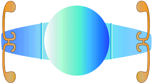
The C & C Vision Crystalens (Slide 6) is a modified plate-haptic lens manufactured from a high refractive index (RI =
1.430), third-generation nonreflective silicone material
(Biosil), which contains an ultraviolet (UV) filter. The lens is hinged
adjacent to the optic and has small looped polyimide haptics,
which have been shown to fixate firmly in the capsular bag.32
The grooves across the plates adjacent to the optic make the
junction of the optic with the plate haptic the most flexible part
of the haptic/optic design. The overall length of the lens is 11.5
mm (loop tip to loop tip measurement), while the overall length
as measured from the ends of the plate haptics is 10.5 mm. The
optic is biconvex and is 4.5 mm in diameter. The A-constant is
119.0, and the lens is designed for placement into the capsular bag.
Preclinical Studies
Extensive preclinical testing of the Crystalens was conducted in
accordance with internationally-recognized standards for
permanent implants. As shown in Table 1, the
Crystalens
intraocular lens met all requirements for biocompatibility of
medical devices.
In addition, the Crystalens was subjected to rigorous optical and mechanical testing in accordance with the guidelines of the U.S. Food and Drug Administration (FDA) for intraocular lenses. In each test, 10 sterile, finished lenses each of low (18.5 D), mid-range (23 D), and high diopter (25 D) were evaluated.
Compression force at 10 mm (immediate and after decay), axial displacement (immediate and after force decay), and angle of contact were all characterized for the Crystalens lens in accordance with FDA guidelines, as well as internationally recognized standards for intraocular lenses. The results of this testing are displayed in Table 2.
Durability of the Crystalens
The third-generation silicone material (Biosil) used in the
manufacturing of the Crystalens has been extensively evaluated to
determine its ability to withstand the multiple flexions that will
result from the repeated changes of focus during normal
functioning of an eye implanted with this lens. A movement of 1
mm can provide up to 2 D of added power, which is
equivalent to the power of a standard bifocal in a pair of glasses.
The number and extent of accommodative cycles the eye
experiences during the course of a day is difficult to estimate.
However, in an analysis conducted by Arthur P. Little, it was
estimated that the average frequency of accommodation is
probably on the order of 20 to 120 per hour (1 cycle every 3
minutes to one every 30 seconds). Assuming that an average
person is awake 16 hours per day, the range would be from 20
cycles per hour (120,000 per year) to 120 cycles per hour
(720,000 per year). With a fatigue lifetime exceeding 20 million
cycles, it has been roughly estimated that the Crystalens should
last a minimum of 30 years and probably much longer.
Clinical Experience With the Crystalens
Initial Safety Studies
Based on the experience acquired with the previous designs
of the accommodating intraocular lens, two further designs
were evaluated. In February 1998, a comparative trial of the two
models, the sixth design, Crystalens model AP-45, and
seventh design, Crystalens model AT-45, was initiated. The design objective was
to eliminate the lens dislocations reported with the previous
designs while providing distance vision and an adequate
accommodative amplitude to perform near and intermediate tasks.
In this comparative trial, patients underwent standard cataract surgery with capsulorrhexis and phacoemulsification. The lens was implanted without folding through a 3.5-mm to 3.7-mm incision. Use of atropine for a period of 3 weeks following surgery was initially utilized to ensure that the lens was fixated in the most posterior position for distance vision and to provide perfect centration and stability. Since atropine paralyzes the ciliary muscle for 2 weeks, the muscle was paralyzed for 5 weeks. The patients were followed for 6 months with the objective of monitoring for dislocation and decentration.
This clinical trial was conducted at three sites: Professor Jochen Kammann, Dortmund, Germany; Rodolfo Benavidez, MD, Monterrey, Mexico; and Ceclio Velasco, MD, Mexico City. A total of 47 patients were enrolled over a 5-month period. Four of the 25 model AP-45 lenses dislocated early in the clinical trial, resulting in discontinued use of this lens. In contrast, there were no dislocations or complications observed in the 22 eyes that received the Crystalens.
Initial Efficacy Studies
Upon resolving the fixation problems associated with the
previous six designs, C & C Vision initiated a second clinical
trial with the Crystalens in Tijuana, Mexico under the direction
of Dr. Arturo Chayet. Chayet is an internationally recognized
ophthalmic surgeon and a leader in the development of new
technologies. Chayet initially began using the Crystalens in
patients whose visual potential could not be evaluated because
dense cataracts prevented observation of the retina. After
conducting his own safety study, Chayet proceeded to
implant the Crystalens lens in younger patients with the
capability of excellent vision postoperatively. In this study, four
measurements of visual acuity have been performed on each
patient, at all examinations, as follows:
- UDVA – Uncorrected distance visual acuity
- UNVA – Uncorrected near visual acuity at 40 cm (16 inches)
- BCDVA – Best-corrected distance visual acuity
- DCNA – Distance corrected near acuity
Distance corrected near acuity is the patient’s ability to read a near target placed 40 cm (14 inches) away from behind the phoropter through the distance correction. This was measured by the ability of the patient to read a Jaeger reading chart. Since the patient’s correction at the phoropter is set for distance, the reading of a near target requires the intraocular lens to adjust for near, thereby suggesting accommodation of the Crystalens. The factors responsible for pseudoaccommodation (i.e., myopia, cylinder, and small pupil) are eliminated by this method of testing near vision. This “accommodating” feature is unique to the Crystalens.
Between January 1999 and February 2000, Dr. Chayet implanted 33 Crystalenses in 19 patients. Twenty-eight of these lenses were implanted in both eyes of 14 patients. The five remaining patients received a Crystalens in one eye only. Bilateral results on 14 patients (28 eyes) are graphically depicted in Slide 7. The bilateral patients implanted had a mean follow-up of 2 years.
Mean intraocular pressure was 15.5 mm Hg postoperatively in these 33 eyes, with a range of 11 mm Hg to 19 mm Hg. No corneal edema or iritis was reported past 1 day postoperatively with the exception of one patient whose mild iritis resolved after 2 weeks, and there were no lens decentrations or dislocations. No complications or adverse events have been reported at any time during the course of the study, to date.
With modern standard cataract surgery utilizing careful biometry, the majority of patients can see 20/40 or better without glasses at distance and some patients can read J3 or better. It is important to note that possible conditions that aid or enable pseudophakic patients to read without glasses, that is, some degree of myopia, astigmatism, or a small pupil (or a combination of these conditions), were eliminated in this study by examining the study patients behind the phoropter in a dimly lit room and having them read a near reading chart at 14 inches through their distance correction. Chayet’s data indicate that near vision improves and is best evaluated after bilateral implantation.
Although these data are preliminary, in this small population of eyes, the Crystalens provided good best-corrected distance visual acuity, consistent with historical controls published by the FDA, with no loss in letter lines from the preoperative best corrected distance vision. Additionally, all bilaterally implanted patients had excellent uncorrected distance and near visual acuities, allowing reading without an add.
Clinical Trials of the Crystalens in the United States
Based on the early results of 20 lenses implanted in 16
patients at three sites in Mexico and the submission to the
FDA of some
requisite biocompatibility studies in rabbits, C & C Vision
received approval from the FDA to conduct a multicenter clinical
trial of the Crystalens in the United States in March 2000.
Clinical Protocol
In the FDA approved study, patients who were candidates for
cataract removal and intraocular lens implantation were enrolled
at 13 investigational sites participating in this prospective multicenter trial. The first phase of the study consisted of five
investigators who enrolled a total of 100 eyes in 100 patients.
Eight additional investigators are in the process of enrolling the
remainder of the cohort population for a total of 425 implants.
Three hundred eyes with 1 year follow-up are required for
submission of an application for marketing approval. Following
approval, a cohort of 300 eyes must be followed for 3 years to
complete the study.
Patients with cataracts who were candidates for extracapsular cataract extraction by phacoemulsification and implantation of an intraocular lens were eligible for enrollment. Other inclusion criteria included age of 50 years or older, potential visual acuity of 20/30 or better in both eyes, adequate mental capacity to allow the conduct of the extensive visual examination required for the study, and informed consent. Additionally, patients were screened for their ability and willingness to return for scheduled follow-up examinations for a period of 3 years after surgery.
Patients with previous ocular surgery, anterior segment pathology, uncontrolled glaucoma or visual field loss from glaucoma, corneal cylinder of more than 1 D, incomplete or damaged zonules, aniridia, congenital bilateral cataracts, microphthalmos, previous retinal detachment or retinal pathology, diabetic retinopathy, macular degeneration or other degenerative visual disorders, or any other ocular pathology that could reduce vision (other than a cataract) were excluded from enrollment.
Patients meeting these entry criteria underwent an extensive preoperative evaluation, including a comprehensive ophthalmic examination, which included slit lamp biomicroscopy, Goldmann tonometry, manifest refraction, and visual acuity measurements. Best-corrected and uncorrected distance acuities were measured using the Stereo Optical 166 Vision Tester (Stereo Optical Inc., Chicago, Ill). Best-corrected near visual acuity was measured pre- and postoperatively using trial frames at 16 inches, using an MN reading card (Lighthouse), which was recommended by FDA for this trial. Near and intermediate acuity at 16 and 32 inches were also measured uncorrected and through the distance correction and then the distance corrected near vision with an add.
All cases were performed through a clear corneal or scleral tunnel incision of no more than 4 mm in width, using capsulorrhexis and phacoemulsification; no sutures were used unless deemed medically necessary. Viscoelastic use was limited to a list of commercially available materials. No folding instruments were used and the lens was inserted flat.
Intraoperative medications, as well as postoperative medications (topical steroid and antibiotic) of the surgeon’s choice, were utilized, with each site adhering to a standardized perioperative regimen. All patients received treatment with atropine at the end of surgery and on the first postoperative day, with the intent of paralyzing the ciliary muscle to allow fixation of the Crystalens in a posterior distance vision position during fibrosis of the capsular bag during the initial postoperative period.
Clinical Results
Patient Demographics
Demographic information is provided in Table 3 for the 50 eyes
in Phase 1 of the FDA Study that have reached 1 month or
more postoperative follow-up. The study cohort was composed
of eyes from patients with a mean age of 71.6 years (range 50
to
88 years). More than half of all patients were men, and the remaining
of the patients were women. At baseline, all patients had normal
slit lamp examinations, other than the presence of a cataract, and
normal intraocular pressures. None of the qualified patients
were excluded from enrollment based on exclusions at the time
of surgery, such that all screened patients were implanted with
the Crystalens.
Efficacy Results
It is important to emphasize that accurate biometry is essential to enable patients to see at all distances without glasses. The biometry in the early cases was performed by applanation, and optical coherence using automatic K readings. Lens power selections and postoperative refractions were reviewed by the clinical staff of C & C Vision and the lens power selected in collaboration with the clinical investigator. After carefully evaluating the different methods of biometry, it was determined that the best results were obtained using immersion biometry with manual K readings, which was subsequently used for the remainder of the study. A lens power to provide a postoperative refraction of close to –0.50 was selected for the first eye and plano will be selected for the second eye in Phase 2 of the FDA study, provided that the refraction in the first eye was on the minus side.
Slide 8 contains the results from the 50 eyes that were implanted with the Crystalens. 48 eyes had a corrected distance vision of 20/25 or better, of these the intermediate vision was excellent and 48/50 (85%) could read J3 or better through their distance correction.
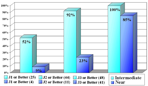
The data presented in Slide 8 show that excellent distance corrected near and intermediate visual acuities are achieved in the majority of patients implanted with a Crystalens. Furthermore, while the 50 eye cohort represents a small sample of the total study population, the data suggest that patients continue to have excellent uncorrected near, intermediate, and distance visual acuities with the Crystalens.
Safety Results
No flare or cells, iritis or corneal edema were reported after
2
weeks postoperatively.
Summary and Conclusions
The results of this initial phase of study of the
accommodating lens suggest that good uncorrected near,
intermediate and distance visual acuity can be achieved
with the appropriate lens design in postoperative
cataract patients. Phase 2 of the FDA
Clinical trial is currently under study.
Issues and Concerns
Complications
Breakthrough technology is rarely introduced without clinical
challenges. As previously discussed, the early models of the
accommodating intraocular lens were associated with
unacceptable rates of dislocations and decentrations. As a result
of several important design changes, no further complications or
adverse events have been associated with the Crystalens design.
Centration has been excellent, there have been no dislocations,
and the ability of the patients to see at near and intermediate
through their distance correction has been variable but observed
consistently in all patients, resulting in excellent patient
satisfaction in more than 800 implants worldwide. Seventeen
YAG capsulotomies have been reported, with no effect on
the ability of these patients to see at near through their distance
correction post-YAG.
Despite the small optic size (4.5 mm), there have been no significant reports of glare with any of the accommodating intraocular lens designs studied. This is probably due to three factors: 1) the deep placement of the optic within the eye, 2) excellent centration, and 3) the non-reflective nature of the third-generation silicone material. Stephen Slade, MD, the medical monitor for the C & C Vision U.S. multicenter clinical trial of the Crystalens, postulates that the lens is so retroplaced, its 4.5-mm optic is actually equivalent to a 5.8-mm or 5.9-mm optic of a lens located in the anterior capsular bag space.
Glare and contrast sensitivity were tested in a comparative study reported at the 2000 American Society for Cataract and Refractive Surgery (ASCRS) meeting in San Diego, California, conducted by Michael Colvard, MD, one of the principal investigators in the U.S. multicenter clinical trial of the Crystalens. Glare and contrast sensitivity were measured in 10 Crystalens implanted eyes (mean age = 75.7 years), 10 eyes (average age = 72 years) implanted with a silicone multifocal intraocular lens with a 6-mm optic, and 10 eyes (mean age = 75.3 years) implanted with a 6-mm monofocal acrylic intraocular lens. The mean follow-up was 1 year. Test results were independently analyzed by leading contrast vision expert, Arthur Ginsburg, PhD, of the Vision Science Research Group in San Ramon, California.
Colvard noted that although the small sample size precludes definitive conclusions, the results from this study indicated that neither the smaller optic nor the third generation silicone material (Biosil) in the Crystalens increases glare symptoms nor causes loss of contrast sensitivity.
Long-term Safety and Effectiveness
Although results of ongoing clinical trials are still preliminary,
visual performance following implantation of the
Crystalens
has been excellent. As previously reported, in the 11 eyes
(average patient age = 80 years) implanted by Kammann with the second design, distance and near vision
remain unchanged from previous examinations 3½ years after
implantation and all patients read J3 or better through their
distance correction. A-scans on 11 of these eyes before and after
pilocarpine demonstrated anterior movement of the optic of
approximately 1 mm, 12 months postoperatively.
In the multicenter clinical trials conducted in Mexico, 14 bilateral patients (average age = 71 years) implanted with the Crystalens by Arturo Chayet, MD, 100% (14/14) of the patients are seeing 20/40 or better uncorrected distance and J3 or better at near, 70% (11/14) had 20/30 or better uncorrected distance and J3 or better at near, 2 years after surgery.
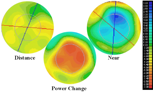
In the U.S. multicenter clinical trials, 98% (41/42) of the patients are seeing 20/25 or better with their corrected distance vision and 93% (39/42) see J3 or better through their distance correction at near, 11 months or longer after surgery. In addition, A-scans have demonstrated anterior movement of the optic and retinal wavefront power maps have shown power changes of up to 3 D (Slide 9).
Conclusion
While several approaches have been used with reasonable success to provide near vision in pseudophakic patients, none of the current modalities utilize the natural physiology of the ciliary muscle following cataract removal. The C & C Vision intraocular lens was specifically designed to allow the lens to move forward to provide near and intermediate vision, upon ciliary muscle constriction.
Irrespective of the possible mechanistic basis for the translational movement of the accommodating intraocular lens in the eye, the early clinical findings following lens implantation show that an intraocular lens designed to move forward and backward upon ciliary muscle constriction and relaxation provides substantial functional distance, intermediate and near vision in pseudophakic patients. Subsequent clinical studies on the C & C Vision Crystalens have demonstrated excellent near and intermediate visual acuity measured through the distance correction without an add. In nearly all implanted eyes, as expected, the uncorrected and distance corrected near acuity improved substantially in these cataract patients. Uncorrected distance visual acuities were also excellent, with more than 90% of patients having distance acuities of 20/40 or better.
Based on the extensive body of clinical data generated by more than 30 ophthalmic surgeons who have implanted more than 800 lenses worldwide, the Crystalens has provided excellent uncorrected vision for both distance, intermediate, and near in the majority of patients, reducing their need for spectacle correction.
References
- Glasser A, Campbell MCW. Presbyopia and the optical changes in the human crystalline lens with age. Vision Res 1998;38(2):209-229.
- Glasser A, Campbell MCW. Biometric, optical and physical changes in the isolated human crystalline lens with age in relation to presbyopia. Vision Res. 1999;39:1991-2015.
- Schachar RA, Bax AJ. Mechanism of human accommodation as analyzed by nonlinear finite elemental analysis. Compr Ther. 2001;27(2):122-132.
- Nishi O, Nakai Y, Yamagda Y, Mizumoto Y. Amplitudes of accommodation of primate lenses refilled with two types of inflatable endocapsular balloons. Arch Ophthalmol. 1993;111:1677-1684.
- Hara T, Sakka Y, Sakanishi K, et al. Complications associated with endocapsular balloon implantation in rabbit eyes. J Cataract Refract Surg. 1994;20:507-512.
- Parel J, Gelender H, Trefers W, Norton E. Phaco-ersatz: cataract surgery designed to preserve accommodation. Graefes Arch Clin Exp Ophthalmol. 1986;224:165-173.
- Haefliger E, Parel J, Fantes F, Norton E, Anderson D, Forster R, Hernandez E, Feuer W. Accommodation of an endocapsular silicone lens (Phako-Ersatz) in the nonhuman primate. Ophthalmology. 1987;94:471-477.
- Fisher R. The force of contraction of the human ciliary muscle during accommodation. J Physiol. 1977;270:51-74.
- Fisher R. The mechanics of accommodation in relation to presbyopia. In: Stark L, Obrecht G, eds. Recent Research and Reviews from the Third International Symposium. New York: Professional Press; 1987:42-44.
- Fisher R. The mechanics of accommodation in relation to presbyopia. Eye. 1988;2:646-649.
- Fisher R. The ciliary body in accommodation. Trans Ophthalmol Soc UK. 1986;105:208-219.
- Glasser A, Kaufman PL. The mechanism of accommodation in primates. Ophthalmolology. 1999;106: 863-872 .
- Bito L, DeRousseau C, Kaufmann P, Bito J. Age dependent loss of accommodative amplitude in rhesus monkeys: An animal model for presbyopia. Invest Ophthalmol Vis Sci. 1982;23:23-31.
- Haefliger E, Parel J. Accommodation of an endocapsular silicone lens (Phako-Ersatz) in the aging rhesus monkey. J Ref Corneal Surg. 1994;10:550-555.
- Strenk SA, Semmlow JL, Strenk LM, Munoz P, Gronlund- Jacob J, DeMarco JK. Age-related changes in human ciliary muscle and lens: A magnetic resonance imaging study. Invest Ophthalmol Vis Sci. 1999;40(6): 1162-1169.
- Thornton S. Lens implantation with restored accommodation. Current Canadian Ophthalmic Practice. 1986;4:60-62.
- Thornton S. Accommodation in pseudophakia. Color Atlas of Lens Implantation. 1991;159-162.
- Huber C. Planned myopic astigmatism as a substitute for accommodation in pseudophakia. Am Intraocular Implant Soc 1981;7:244.
- Huber C. Myopic astigmatism as a substitute for accommodation in pseudophakia. Doc Ophthalmol. 1981;52:123.
- Nakazawa N, Ohtsuki K. Apparent accommodation in pseudophakic eyes after implantation of posterior chamber intraocular lenses. Am J Ophthalmol. 1983;96:435-438.
- Trindade F, Oliveira A, Frasson M. Benefit of against-the-rule astigmatism to uncorrected near acuity. J Cataract Refract Surg. 1997;23:82-85.
- Percival S. Prospectively randomized trial comparing the pseudoaccommodation of the AMO ARRAY multifocal lens and a monofocal lens. J Cataract Refract Surg. 1993;19:26-31.
- Busacca A. La physiologie du muscle ciliaire etudiee par la gonioscopie. Annales D’Oculistique 1955;1-21.
- Coleman J. On the hydraulic suspension theory of accommodation. Trans Am Ophthalmol Soc. 1986;846-868.
- Hardman Lea S, Rubinstein M, Snead M, Haworth S. Pseudophakic accommodation? A study of the stability of capsular bag supported, one piece, rigid tripod, or soft flexible implants. Br J Ophthalmol. 1990;74:22-25.
- Niessen A, De Jong L, Van der Heijde G. Pseudoaccommodation in pseudophakia. Eur J Implant Refract Surg. 1992;4:91-94.
- Cumming JS, Ritter J. The measurement of vitreous cavity length and its comparison pre- and postoperatively. Eur J Implant Refract Surg. 1994;6:261-272.
- Colin, J. Clinical results of implanting a silicone hapticanchor-plate intraocular lens. J Cataract Refract Surg. 1996;2:1286-1290.
- Cumming JS. Postoperative complications and uncorrected acuities after implantation of plate haptic silicone and three-piece silicone intraocular lenses. J Cataract Refract Surg. 1997;19:263-274.
- Strenk SA. Age-related changes in human ciliary muscle and lens: A magnetic resonance imaging study. Invest Ophthalmol Vis Sci. 1999;40(6).
- Kammann J. Vitreous-stabilizing, single-piece, mini-loop, plate-haptic silicone intraocular lens. J Cataract Refract Surg. 1998;24:98-106.
- Kent D, Peng Q, Isaacs R, Whiteside S, Barker D, Apple D. Mini-haptics to improve capsular fixation of plate haptic silicone intraocular lenses. J Cataract Refract Surg. 1998;24:666-671.